From Nashiji to Peau d'Orange: A Japanese Story Behind Angioid Streaks
Published in General & Internal Medicine and Arts & Humanities
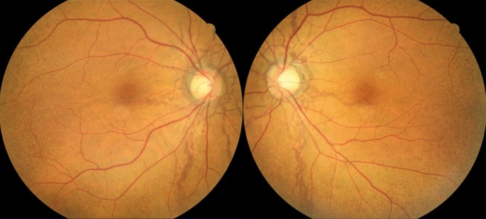

Our study began with a clinical question. We wanted to clarify the fundus findings in Japanese patients with angioid streaks and to examine their association with pseudoxanthoma elasticum (PXE). During this work, we became interested in the history of the term Nashiji fundus.
Peau d’orange is recognized as an important ocular finding of PXE. It describes a mottled appearance of the fundus resembling the surface of an orange peel. In our study, peau d’orange was observed more frequently than clinically diagnosed PXE. This finding suggested that PXE may not have been fully recognized in some patients with angioid streaks.
In Japan, this fundus appearance has also been called Nashiji fundus. Nashiji refers to the finely speckled surface of a Japanese pear. The same word is used for Nashiji maki-e, a traditional Japanese lacquer technique in which fine metallic particles are applied to produce a granular appearance similar to pear skin. The term therefore gives Japanese ophthalmologists a familiar image of this characteristic fundus pattern.
Professor Koichi Shimizu1 reported this finding in a Japanese article titled “Nashiji fundus in pseudoxanthoma elasticum” in 1960. In the following year, he published an English-language article titled “Mottled fundus in association with pseudoxanthoma elasticum” in the Japanese Journal of Ophthalmology.2 The Japanese term Nashijiwas translated as “mottled fundus” and did not become widely known outside Japan. Several years later, Smith and Gass3 used the term peau d’orange, which subsequently became established in the international ophthalmic literature.
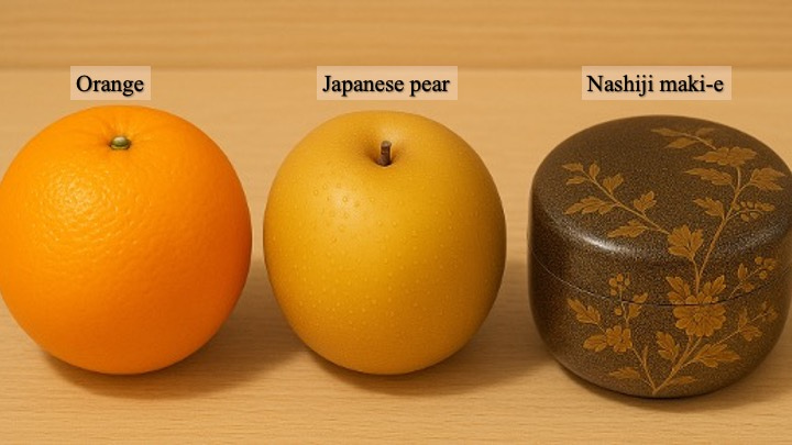
The metaphor may change, but the mottled pattern remains.
Nashiji fundus is still used in Japanese ophthalmology. Japanese textbooks and clinical teaching materials often present it together with the international term, as Nashiji fundus (peau d’orange) or 梨子地眼底. Thus, both terms remain familiar to Japanese ophthalmologists.
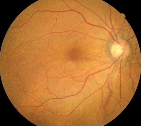
This Japanese term"梨子地眼底" is still used in Japanese textbooks.
Our study, “Clinical characteristics of angioid streaks in Japanese patients,” included 33 patients. PXE was clinically diagnosed in 45.5% of the patients, whereas peau d’orange was observed in 54.5% of the eyes. Choroidal neovascularization and macular atrophy were each found in approximately half of the eyes. We also evaluated comet tail lesions, pattern dystrophy-like changes, subretinal drusenoid deposits, outer retinal tubulation, and optic nerve head drusen.
The difference between the frequency of clinically diagnosed PXE and that of peau d’orange was an important finding. PXE may be difficult to diagnose when its skin manifestations are mild or have not been examined histologically or genetically. In such cases, characteristic ocular findings may provide a clue to the presence of systemic disease. Therefore, patients with angioid streaks should be carefully examined for peau d’orange and other findings associated with PXE.
Our results also showed that angioid streaks are associated with various retinal and choroidal changes. The high frequency of choroidal neovascularization indicates the need for long-term follow-up. Macular atrophy, photoreceptor-related structural changes, and choroidal thinning may reflect progressive changes occurring over the course of the disease.
By revisiting the term Nashiji, we do not intend to replace the internationally accepted term peau d’orange. Rather, we hope to introduce the historical and cultural background of the Japanese terminology. The same fundus finding has been described using different visual references: orange peel in the international literature and Japanese pear skin in Japan.
This study allowed us to connect current clinical imaging with earlier observations made by Japanese ophthalmologists. We hope that our findings will encourage careful examination for PXE-related ocular signs in patients with angioid streaks and will also bring renewed attention to the term Nashiji fundus and its place in the history of ophthalmology.
References
1. Shimizu K. Nashiji fundus in pseudoxanthoma elasticum. Jpn Rev Clini Ophthalmol. 1960;54:503–9 (in Japanese).
2. Shimizu K. Mottled fundus in association with pseudoxanthoma elasticum. Jpn J Ophthalmol. 1961;5:1–13.
3. Smith JL, Gass JD, Justice J Jr. fluorescein fundus photography of angioid streaks. Br J Ophthalmol. 1964;48:517–21.


Please sign in or register for FREE
If you are a registered user on Research Communities by Springer Nature, please sign in