Pathways Through Which Iron Deficiency Under HIF-PH Inhibitor Administration Triggers Thrombosis and How to Handle It: A New Mechanism Proposal from the Six-Factor Theory of Erythropoiesis Control
Atsuo Takemasa, Nishinokyo Hospital
Regarding the mechanism by which iron deficiency under HIF-PH inhibitor administration promotes thrombosis, it has traditionally been thought that an increase in transferrin (Tf) is the cause. However, the Tf gene does not have an IRE, so the increase in Tf is HIF-1α dependent (Fig. 3, ㉒). Tf itself is not a coagulation factor, but it can bind to thrombin and FXIIa and enhance their activity as a coagulation-promoting effect. It is also thought to interfere with antithrombin's inhibition of thrombin 1,2). On the other hand, there are increasing reports suggesting that Tf might actually help prevent thrombosis 3,4). One proposed mechanism is that Tf increases its iron-binding capacity and reduces free iron, thereby reducing oxidative stress and vascular damage, and acting against thrombosis, though this is not fully investigated. Also, Tf levels in the blood can fluctuate significantly due to inflammation or changes in iron metabolism. Therefore, depending on the situation, it could either promote or inhibit coagulation. In conclusion, while Tf may serve as one indicator of thrombosis risk when using HIF-PH inhibitors, it’s probably wrong to say it’s the main cause of thrombosis.
TFPI (Tissue Factor Pathway Inhibitor) is a Kunitz-type protease inhibitor that specifically inhibits FXa and the TF-FVIIa complex, mainly affecting the initiation of coagulation. TFPIα is considered the most important pool of TFPI when it comes to changing the risk of thrombosis or bleeding. In plasma, TFPIα forms a ternary complex with FV-short and protein S. The expression level of FV-short controls circulating TFPIα levels, and protein S enhances anticoagulant function through its essential cofactor 5). On the other hand, HIF-1α and TNF-α suppress TFPI 6,7), and the ratio of Tissue Factor (TF) to TFPI becomes a key determinant of FX activation 8).
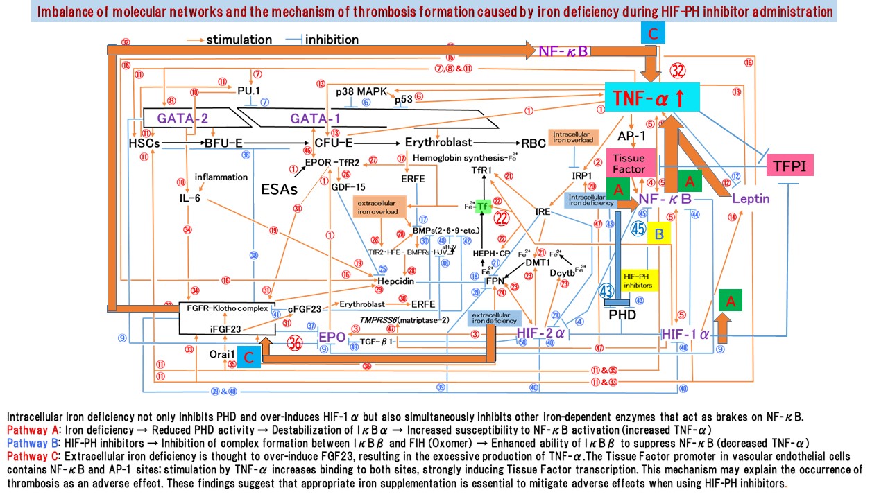
Based on the above, I'm reporting that, using the 'Six-factor theory of erythropoiesis control' as a new mechanism, I theoretically discovered the pathway of thrombosis. I modified Figure 1 from the Six-factor theory and show it as Figure 3. Intracellular iron deficiency not only inhibits PHD (Fig. 3, ㊸) and excessively induces HIF-1α, but also simultaneously suppresses other iron-dependent enzymes that act as brakes for NF-κB.
Pathway A: Intracellular iron deficiency → reduced PHD activity → IκBα destabilization → increased susceptibility to NF-κB activation → upregulation of TNF-α. PHD is an enzyme that requires ferrous iron (Fe²⁺) at its active site. Iron chelation strips iron from PHD, causing a marked decline in PHD activity even in the presence of sufficient oxygen. This abolishes the PHD-mediated hydroxylation of proline residues on IKKβ (an inhibitory signal). Consequently, IKKβ becomes active, leading to the phosphorylation, ubiquitination, and subsequent degradation (destabilization) of IκBα. The released NF-κB (p65/p50) translocates into the nucleus and directly upregulates TNF-α gene expression 9, 10).
Pathway B: HIF-PH inhibitors → inhibition of complex formation between IκBβ and FIH (Oxomer) → increased ability of IκBβ to suppress NF-κB → suppression of TNF-α(Fig. 3, ㊺). The primary function of IκBβ is to bind to NF-κB transcription factor dimers—specifically those containing c-Rel and p65—and sequester them in the cytoplasm, thereby preventing gene expression driven by these specific NF-κB dimers. When IκBβ binds to FIH to form an Oxomer, it loses its ability to interact with the NF-κB subunits p65 and c-Rel. Oxomer formation promotes NF-κB-dependent transcription by inhibiting the binding of p65 and c-Rel to IκBβ. Pathway B represents one aspect of the pharmacological action of HIF-PH inhibitors; it is thought that, provided iron levels are sufficient, a balance can be maintained between Pathway B and Pathway A 11,12).
Pathway C: Extracellular iron deficiency excessively induces FGF23 (Fig. 3, ㊱), resulting in the excessive production of TNF-α (Fig. 3, ㉜). Figure 3 illustrates pathways A through C.
TF and TNF-α mutually potentiate each other. TNF-α strongly induces TF expression; subsequently, the induction of TF leads to the generation of thrombin and FXa—byproducts of the coagulation cascade—which activate NF-κB and further increase TNF-α levels, thereby establishing a vicious cycle. FXa activates NF-κB via PAR-2, while thrombin activates it via PAR-1 13, 14, 15). Additionally, it has been suggested that FGF23 may impair endothelial function through the activation of NF-κB 16).
TNF-α simultaneously activates MAPK pathways—such as JNK—and AP-1, a complex composed of c-Jun and c-Fos. TNFR1 rapidly activates the IKK system (NF-κB). The TF promoter in vascular endothelial cells contains both NF-κB and AP-1 binding sites; TNF-α stimulation increases binding at both sites, strongly inducing TF transcription. NF-κB acts as the "initial switch." In contrast, the MAPK system (AP-1) functions as an "amplifier"—acting with a delay due to its long kinase cascade—to powerfully boost TF promoter activity initiated by NF-κB17, 18). Sustained TF expression serves as a molecular trigger for the development of thrombosis. A distinctive feature of the iron-deficiency stress caused by HIF-PH inhibitors is the rapid onset of intracellular iron depletion, which is more severe than that seen in conventional iron deficiency. Although cells attempt to aggressively take up iron, the supply fails to keep pace, resulting in marked intracellular iron depletion that readily triggers NF-κB activation and subsequent TNF-α induction (Pathway A). In conclusion, rather than viewing increased Tf levels as the primary cause of thrombosis, it is more consistent with the data—and clarifies potential intervention points—to reinterpret the phenomenon as a network centered on the HIF/NF-κB/TNF-α axis, the FGF23/NF-κB/TNF-α axis, and the iron deficiency/NF-κB/TNF-α/TF-TFPI axis.
The "Six-factor theory of erythropoiesis control" has provided a theoretical explanation for the mechanism underlying thrombosis—a side effect of HIF-PH inhibitors observed during iron deficiency. As a plan to validate this new theory, we intend to begin by measuring blood levels of TF and TFPI during HIF-PH inhibitor administration. When using HIF-PH inhibitors, the key to preventing thrombosis lies in avoiding unmanaged iron deficiency and ensuring appropriate iron supplementation. Thrombosis is likely not the only side effect that can be prevented. By conceptualizing erythropoiesis as a system, the "Six-factor theory of erythropoiesis control" successfully incorporates the coagulation-fibrinolysis system into its network—extending beyond erythropoiesis, infection defense, bone metabolism, and iron metabolism.
References
1) Tang, X. et al. Transferrin plays a central role in coagulation balance by interacting with clotting factors. Cell Res. 30, 119–132 (2020).
2) Tang, X. et al. Iron-deficiency and estrogen are associated with ischemic stroke by up-regulating transferrin to induce hypercoagulability. Circ. Res. 127, 651-663(2020).
3) Shah, B.N. et al. Increased transferrin protects from thrombosis in Chuvash erythrocytosis. Am. J. Hematol. 98, 1532-1539 (2023).
4)Ricardo, A. et al. Increased Transferrin Concentrations Are Not Associated With Thrombosis in People Living at High Altitude. J. Hematol. 14, 20-25(2025).
5) Ahnström, J. et al. Tissue factor pathway inhibitor – cofactor-dependent regulation of the initiation of coagulation. Curr. Opin. Hematol. 31, 315-320 (2024).
6) Cui, X.Y. et al. Effect of hypoxia on tissue factor pathway inhibitor expression in breast cancer. J. Thromb. Haemost. 14, 387-396 (2016).
7) Shimokawa, T. et al. Down-regulation of murine tissue factor pathway inhibitor mRNA by endotoxin and tumor necrosis factor-alpha in vitro and in vivo. Thromb. Res. 100, 211-221 (2000).
8) Brandtner, A.K. et al. Differential procoagulatory response of microvascular, arterial and venous endothelial cells upon inflammation in vitro. Thromb. Res. 205, 70-80 (2021).
9) Cummins, E. P. et al. Prolyl hydroxylase-1 negatively regulates IκB kinase-β, giving insight into hypoxia-induced NFκB activity. PNAS. 103, 18154-18159 (2006).
10) Scholz, C.C. et al. Hydroxylase-dependent regulation of the NF-κB pathway. Biol. Chem. 394, 479– 493 (2013).
11) Takeda, K. et al. Inhibition of prolyl hydroxylase domain-containing protein suppressed lipopolysaccharide-induced TNF-α expression. Arterioscler. Thromb. Vasc. Biol. 29, 2132– 2137 (2009).
12) Volkova, Y. L. et al. Selective hypoxia-sensitive oxomer formation by FIH prevents binding of the NF-κB inhibitor IκBβ to NF-κB subunits. Mol. Cell. Biol. 44, 138-148 (2024).
13) Camerer, E. et al. Tissue factor- and factor X-dependent activation of protease-activated receptor 2 by factor VIIa. PNAS. 97, 5255-5260 (2000).
14) Hara, T. et al. Emerging roles of protease-activated receptors in cardiometabolic disorders. J. Cardiol. 81, 337-346 (2023).
15) Rahman, A. et al. Gαq and Gβγ Regulate PAR-1 signaling of thrombin-induced NF-κB activation and ICAM-1 transcription in endothelial cells. Circ. Res. 91, 398-405(2002).
16) Guo, L. et al. Elevated fibroblast growth factor 23 impairs endothelial function through the NF‑κB signaling pathway. J. Atheroscler. Thromb. 30, 138-149 (2023).
17) Parry, G.C. et al. Transcriptional regulation of tissue factor gene expression in human endothelial cells.
Arterioscler. Thromb. Vasc. Biol. 15, 612-621(1995).
18) Fujioka, S. et al. NF-κB and AP-1 Connection: Mechanism of NF-κB-Dependent Regulation of AP-1 Activity. Mol. Cell. Biol. 24, 7806-7819(2004).
Keywords: Erythropoietin, Tumor Necrosis Factor-α, Iron Deficiency, Thrombosis, Transferrin, Hypoxia-Inducible Factor, Tissue Factor, Tissue Factor Pathway Inhibitor, Nuclear Factor-κB, Hypoxia-Inducible Factor Prolyl Hydroxylase Inhibitors, FIH, IκBα, IκBβ, AP‑1, Fibroblast Growth Factor 23, Renal Anemia, Chronic Kidney Disease