Looking for cancer stem cells signature in renal cancer cells
Published in Cancer

Clear cell renal cell carcinoma is the most common kidney cancer. Prognosis for this type of cancer is generally poor since it is largely resistant to chemo- and radiotherapy. Many studies suggested that cancer stem cells (also called tumor initiating cells) are responsible for development of tumor, disease progression, aggressiveness, metastasis and drug resistance. However, tumorigenic potential of cancer stem cells isolated from established renal cancer cell lines has never been investigated in vivo. Moreover, there are several things that we don’t know about cancer stem cells in clear cell renal cell carcinoma, eg.:
- How to recognize them? Which molecular markers do they display? Can they be characterized by co-expression (or absence) of several markers?
- How do they behave in vivo (e.g. in experimental animals)?
- Can we propose imaging biomarkers for cancer stem cells or cancer enriched with cancer stem cells?
Our study was designed to answer these questions.
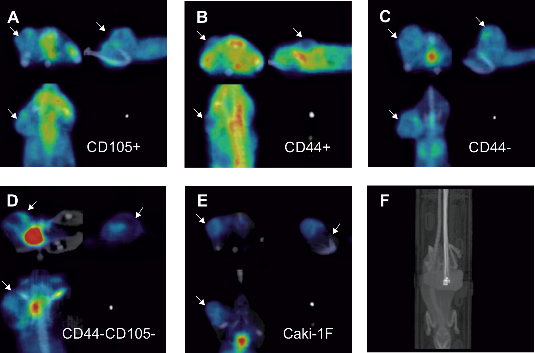
As biomarkers of cancer stem cells multiple surface proteins have been indicated including: CD105, CD133, CD44, or CXCR4; but the co-expression of multiple membrane markers on clear cell renal cell carcinoma cancer stem cells was not defined. We focused on two of these proteins: CD105 (also called endoglin) that is a membrane glycoprotein, part of the TGF beta receptor complex, involved in cytoskeleton organization and angiogenesis (forming of new blood vessels that is crucial for development of tumors) and CD44 which is a membrane glycoprotein involved in cell–cell interactions, cell adhesion and migration of cells. We isolated CD105 positive (CD105+), CD105 negative (CD105-), CD44+ and CD44- cells from Caki-1 renal cancer cell line. We also isolated ‘double stained’ cells, i.e. negative for both CD44 and CD105 (CD44-/CD105-), positive for both markers (CD44+/CD105+) or negative for CD44 and positive for CD105 (CD44-/CD105+). This part of our study confirmed that multiple subpopulations of stem-related phenotype coexist in stable cell line.
These various subpopulations of cells were injected subcutaneously into NOD SCID mice and tumor growth was monitored with noninvasive imaging techniques: magnetic resonance imaging (MRI) and positron emission tomography combined with computed tomography (PET/CT). Tumor growth was observed after implantation of CD105+, CD44+, CD44-, CD44-/CD105+ and CD44-/CD105- but not CD105- or CD44+/CD105+. Implantation of CD44-/CD105- cells induced tumors that were characterized by different image in MRI scans (more specifically they had longer T1 relaxation time) and distinct metabolic pattern than other tumors. All the tumors were characterized by low uptake of PET tracer - [18F] fluorodeoxyglucose. CD105+ and CD44- tumors expressed cancer stem cells’ markers: Nanog and Oct-4, while CD44- tumors additionally expressed endothelial cell marker - CD31.
Importantly, we have shown that CD44-CD105- subpopulation of Caki-1 cells displayed stem-like phenotype. Tumor induced by this subpopulation show specific features distinct from other tumors induced by the other tested subpopulations like different visibility in MRI scans and metabolic fingerprint. For future research in the renal cancer field co-expression of multiple markers will be crucial to define stem cell signature. Primarily reported CD105 positive cells (CD105+) seem to harbor further subpopulations of different tumorigenic potential.
Follow the Topic
-
Scientific Reports
An open access journal publishing original research from across all areas of the natural sciences, psychology, medicine and engineering.

Related Collections
With Collections, you can get published faster and increase your visibility.
Phytochemicals and health
Publishing Model: Open Access
Deadline: Jul 28, 2026
Health Policy and Systems Research
Publishing Model: Hybrid
Deadline: Jul 14, 2026


Please sign in or register for FREE
If you are a registered user on Research Communities by Springer Nature, please sign in