Neuronal Swelling in Hydrocephalus

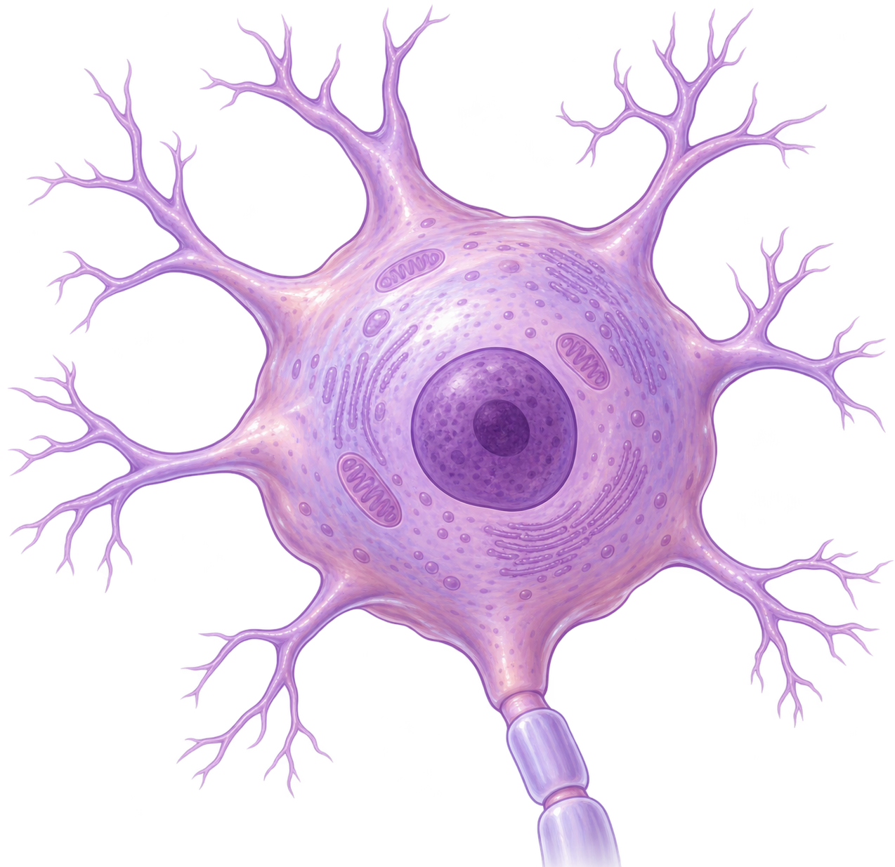
Hydrocephalus is recognized by enlarged brain ventricles, but what happens to individual neurons as the brain is exposed to progressive ventricular expansion? Our study found markedly enlarged neurons in the cingulate cortex of rats with genetic congenital hydrocephalus.
Neuronal Swelling in Hydrocephalus
Like

Share this post

Choose a social network to share with, or copy the URL to share elsewhere

This is a representation of how your post may appear on social media. The actual post will vary between social networks

Explore the Research

BioMed Central
BioMed Central BioMed Central

TRPV4 antagonism attenuates TLR4–NLRP3-mediated neuroinflammation and oxidative stress in cingulate cortex neurons in a genetic rat model of hydrocephalus

Hydrocephalus is characterized by pathological ventricular expansion and periventricular tissue damage, although the molecular mechanisms governing parenchymal injury in this condition remain unclear. Surgical interventions, such as cerebrospinal fluid (CSF) shunting are the standard of care; however, high complication rates underscore the urgent need to understand the underlying pathophysiology and develop targeted pharmacological therapies. Current therapeutic studies focus on reducing CSF secretion, but whether these interventions directly protect brain parenchyma is unknown. Here, using the Tmem67–/– rat model of congenital hydrocephalus, we identified a distinct mechano-inflammatory axis driving cortical neurodegeneration. We found that ventricular distension was accompanied by increased activation of Toll-like receptor 4 (TLR4) and NOD-like receptor family pyrin domain-containing 3 (NLRP3) inflammasomes in cingulate cortex (CgCt) neurons adjacent to the corpus callosum. Inflammatory signaling was localized within neuronal populations, although non-neuronal inflammatory involvement was also observed in the CgCt. Neuronal inflammation was accompanied by mitochondrial oxidative stress-associated signaling in the CgCt and lipid peroxidation, culminating in hypertrophy, and apoptosis. Treatment with transient receptor potential vanilloid 4 (TRPV4) antagonist RN1734 significantly reduced TLR4 and NLRP3 inflammasome expression and lipid peroxidation, although it did not- fully rescue neuronal hypertrophy. Therefore, these findings suggest that TRPV4 antagonism may attenuate hydrocephalus-associated inflammation within cortical tissue by reducing pathological CSF production while simultaneously attenuating downstream inflammatory and oxidative signaling within CgCt.

Hydrocephalus is commonly viewed as a disorder of cerebrospinal fluid (CSF) accumulation and ventricular enlargement. However, as the ventricles progressively expand, the surrounding brain is exposed to substantial mechanical deformation. Structures immediately adjacent to the ventricles, including the corpus callosum and overlying cortex, may therefore be particularly vulnerable to injury. Using a rat model of genetic congenital hydrocephalus, we asked a simple but important question:

What happens to neurons in the cortex surrounding the enlarged ventricles?

We found markedly enlarged neurons

When we examined the pericallosal cingulate cortex, one of the most striking observations was the presence of unusually enlarged neuronal cell bodies in hydrocephalic animals. These neurons were located within regions showing substantial cortical disorganization and compression of surrounding white-matter structures.

To confirm the identity of these cells, we used NeuN, a marker of mature neurons, together with CTIP2, a marker associated with deep-layer cortical neurons. CTIP2 and NeuN showed strong overlap across experimental groups, confirming that the pathological analyses were being performed within a consistent deep-layer neuronal population. Importantly, the enlarged neurons observed in the hydrocephalic cortex were also NeuN+/CTIP2+ cells.

Quantitative analysis showed that the average neuronal soma area increased in hydrocephalic animals, more than a two-fold increase in neuronal size. This pronounced neuronal enlargement or hypertrophy suggests that hydrocephalus affects not only the overall architecture of the brain but also the morphology of individual neurons.

Why is this important?

These findings reinforce an important concept: hydrocephalus affects much more than ventricular size. The expanding ventricles are surrounded by living brain tissue containing neurons, glia, white-matter tracts, and vascular structures. Understanding what happens to these cells as hydrocephalus progresses is therefore essential when evaluating potential therapies.

Link to the full article here: https://doi.org/10.1186/s12974-026-04031-7

Follow the Topic

Neuroscience
Life Sciences > Biological Sciences > Neuroscience
Neurological Disorders
Life Sciences > Health Sciences > Clinical Medicine > Neurology > Neurological Disorders
Neurology
Life Sciences > Health Sciences > Clinical Medicine > Neurology
Neuroanatomy
Life Sciences > Biological Sciences > Neuroscience > Neuroanatomy
Cell Biology
Life Sciences > Biological Sciences > Cell Biology
Spotlight on Research from Japan
Research Publishing > Spotlight on Research from Japan