When cancer hides its origins: unique insights from Barrett's esophagus and esophageal adenocarcinoma

Half of esophageal cancer patients have no Barrett's esophagus at diagnosis. By integrating the epidemiological and multi-modal longitudinal genomic data of 3,100 patients, we found one universal pathway with Barrett's as the hidden precursor, consumed and overwritten by the tumour before diagnosis.
When cancer hides its origins: unique insights from Barrett's esophagus and esophageal adenocarcinoma
Like

Share this post

Choose a social network to share with, or copy the URL to share elsewhere

This is a representation of how your post may appear on social media. The actual post will vary between social networks

There is a question that has followed esophageal cancer clinicians and researchers for a long time, and it goes something like this: if Barrett's esophagus is the precursor to esophageal adenocarcinoma, the cancer of the food pipe, then why do about half of all esophageal cancer patients have no detectable Barrett's at the time of diagnosis?

We set out to answer it. What we found challenged our own assumptions time and again.

Why this question kept us up at night

Esophageal adenocarcinoma (cancer of the food pipe) is one of the worst-outcome solid tumours, and its incidence has risen sharply in Western countries over the past four decades. The only clinically-established early detection route is surveillance of Barrett's esophagus, an endoscopically visible patch of abnormal lining that is considered the pre-cancerous state. The uncomfortable reality is that almost all patients (>90%)1 with this cancer have never been found to have Barrett's esophagus with the current cancer control approaches. Either we are missing an unknown, independent cancer progression pathway that bypasses Barrett's entirely, or Barrett's is always present, but we simply cannot see it. These two possibilities carry very different consequences for how we screen, clinically monitor, and how we framework cancer early detection.

The centrality of this question meant we needed a large, carefully annotated, and deeply phenotyped cohort of patients with research-grade matching normal, Barrett’s, and tumour biospecimens collected cross-sectionally and longitudinally for next generation sequencing. After two decades of work building the OCCAMS cohort, we thought the data and research conditions were finally satisfactory enough to begin studying this question.

What we did

                                                                                                                                                                                                                                                                      Study overview graphic: Understanding why some esophageal cancers develop without a known warning sign                                                                                                                                                                                                                                                                                                                                                                                                                                  This three-part illustration summarizes a large study into esophageal adenocarcinoma (EAC) — a type of gullet cancer — asking whether it always develops from a pre-cancerous condition called Barrett's esophagus (BE).                                                                                                                                                                                                                                                                                                                Part 1 — How common is "Barrett's-negative" cancer?                                                                                                                                                                                                                 Researchers reviewed records from 3,100 patients diagnosed with EAC. Two independent experts examined each case for signs of Barrett's esophagus. They found that 58% of patients had evidence of Barrett's (BE+ve), but a striking 42% had no such evidence        (BE-ve) — meaning nearly half of cases arose without the usual warning sign that clinicians look for.                                                                                                                                                                                                                                                                                                                                                                                                                                   Part 2 — What makes these two groups different?                                                                                                                                                                                                                     The team compared the two groups using both patient-level information (age, sex, BMI, smoking history, heartburn symptoms, and tumor stage) and molecular data from tumor DNA sequencing — including the overall mutation burden, specific mutation patterns, and    cancer-driver genes. A statistical method called logistic regression was used to weigh all these factors together and identify what distinguishes BE-negative from BE-positive cancers.                                                                                                                                                                                                                                                                                                                                                Part 3 — Why might BE-negative cancers grow faster or be harder to catch?                                                                                                                                                                                           Two possible explanations are explored. First, BE-negative tumors show greater subclonal heterogeneity — their evolutionary family trees are more branched and complex than BE-positive tumors, suggesting more aggressive, faster-evolving cancer cell             populations. Second, among 214 patients with Barrett's esophagus who were under regular clinical surveillance, some still went on to develop BE-negative cancer — so-called interval tumors that arose between surveillance visits, highlighting a potential gap    in current screening strategies.
Our study integrates epidemiological, genomic, and spatial analyses across 3,100 patients with esophageal adenocarcinoma (EAC) and Barrett's esophagus (BE).  Alt text: A schematic diagram showing analysis layers  flowing from a central cohort of 3,100 patients, beginning with a question of whether all EAC arise from BE and concluding with a question of what may explain tumor growth.

Through the OCCAMS consortium,2 a network of 25 UK hospitals, we have prospectively enrolled >4,000 patients with esophageal adenocarcinoma between 2002 and 2022 and included 3,100 in our study. For every single tumor, at least two expert pathologists reviewed an array of clinical and pathological reports, surgical specimens, and microscopy slides to determine whether Barrett's esophagus was present, absent, or unascertainable at the time of cancer diagnosis.

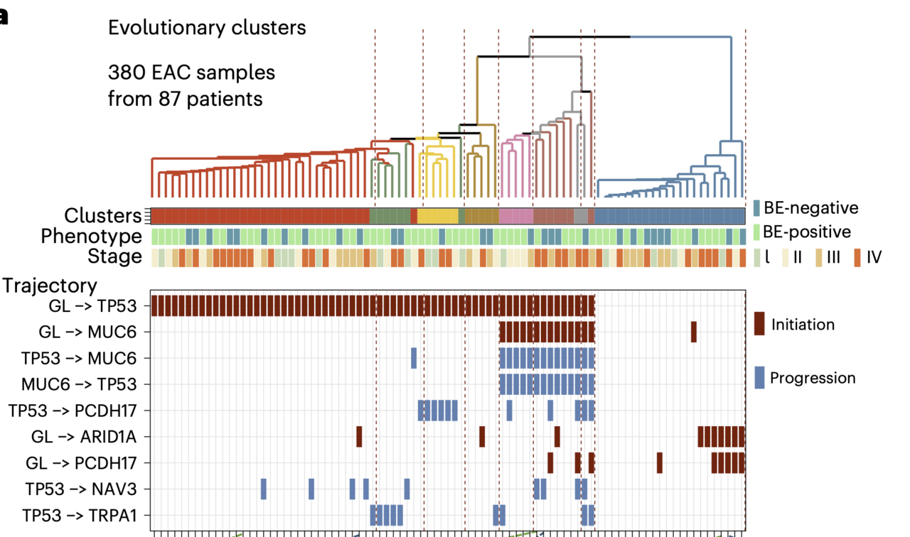
From this cohort, 710 patients had whole-genome sequencing of their tumours basically a complete readout of every DNA change the cancer cells had accumulated. A further 87 patients contributed whole-exome sequencing data from multiple regions within the same tumour, allowing us to reconstruct each cancer's evolutionary history, much like building a family tree. We also assembled a new cohort of 388 cancer-free Barrett's esophagus samples to give us a genomic picture of what the pre-cancerous state actually looks like.

And then, we added one more data layer: spatial transcriptomics that maps gene activity at near-cellular resolution across a tissue section, to ask whether the molecular identity of Barrett's esophagus was retained within tumours, even in patients without any visible Barrett's adjacent to their cancer.

What we found and what surprised us

A three-panel genomic landscape figure comparing Barrett's esophagus (pre-cancer) samples with two types of oesophageal cancer: those arising from Barrett's (BE-positive EAC) and those arising without it (BE-negative EAC). The top section of each panel      shows colour-coded bars representing patient characteristics — including sex, age, smoking history, BMI, and heartburn — alongside dot plots showing each tumour's mutation burden and genomic instability. The bottom section shows which cancer-driver genes      are mutated in each group, and how often, using coloured tiles per patient. Notably, whole-genome doubling (a hallmark of advanced cancer) is rare in pre-cancerous Barrett's tissue (16%) but common in both cancer types (~72–76%). The mutation patterns and     driver gene frequencies look broadly similar between Barrett's-positive and Barrett's-negative cancers, suggesting they share similar genomic mechanisms despite different clinical origins.
Oncoplots comparing clinical and genomic features across the cohort: Barrett's esophagus (BE) samples (n=284 sequencing datasets from 388 BE samples, 256 patients; left), BE-positive EAC (BE+ve EAC; n=252 tumors from 252 patients; centre), and BE-negative EAC (BE-ve EAC; n=183 tumors from 183 patients; right). Upper tracks show patient demographics (sex, age, smoking status, BMI, heartburn) and genomic summary metrics: whole-genome doubling (WGD), tumour mutational burden (TMB; log₁₀mutations/Mb), percentage of aberrant genome (PAG), loss of heterozygosity (LOH), and mutational signature contributions (SBS17a/b). Lower panels show somatic alteration frequencies for 22 recurrently altered cancer driver genes; bar charts on the right of each panel indicate the percentage of samples harbouring each alteration. Alteration types are colour-coded: SNV/indel (green), deletion (blue), amplification (red/pink), and combined events (yellow, purple). Asterisks (*) denote statistically significant differences in the BE cohort. WGD frequency increases markedly from BE (16%) to EAC (72–76%), with broadly similar genomic profiles between BE+ve and BE-ve EAC, though heartburn prevalence is lower in BE-ve EAC (76%) compared to BE (93%) and BE+ve EAC (83%).

When the genomic data came back, the similarity between patients with and without Barrett's esophagus was striking. This was in addition to the remarkable similarity we had already observed given the patient-level epidemiological data analysis. The same genes were mutated at basically the same rates. The so-called mutational signatures, chemical "fingerprints" left in DNA by repeated acid and bile exposure, characteristic of Barrett's esophagus, were equally present in both groups. Tumour evolution also followed the same trajectories. Regardless of how many angles we looked from76 known esophageal cancer-driving genes, copy number changes across every chromosome arm, rare catastrophic genomic events, we could not find a feature that distinguished a "Barrett's-independent" cancer from a Barrett's-associated one.

We next integrated the clinical and molecular data in an adjusted logistic regression model and found the only variable that reliably separated the two groups was tumour stage: patients without Barrett's presented with more advanced cancer.

A forest plot — a standard way researchers display statistical results — showing which tumour and genomic features   are more common in Barrett's-negative oesophageal cancer (BE-ve EAC) compared to Barrett's-positive cancer (BE+ve EAC).    Each row is a different factor; dots represent the estimated effect size (odds ratio) and horizontal lines show the   range of uncertainty (95% confidence interval). Dots to the right of the centre dashed line indicate a stronger link to    BE-negative cancer. Two estimates are shown per factor: one adjusting for patient characteristics (brown) and one   without adjustment (blue).    ▎ The clearest finding is that BE-negative cancers are more likely to be at an advanced stage at diagnosis — T stage   III and IV tumours show odds ratios around 3–4 times higher compared to early-stage (T stage I) tumours. By contrast,   DNA-level features of the tumour — including overall mutation burden, specific mutation patterns (SBS17), whole-genome   doubling, and individual driver gene mutations — show no meaningful difference between the two cancer types, suggesting    the two phenotypes are genomically similar despite their different clinical presentations.
Odds ratios (OR) with 95% confidence intervals for genomic and tumour-stage variables from logistic regression models, with BE-negative EAC as the outcome and BE-positive EAC as the reference. Estimates are shown for unadjusted (blue) and adjusted (red) models; adjusted models account for age, sex, smoking, heartburn, and BMI. The x-axis is log-scaled; the dashed vertical line at OR = 1.0 indicates no association. Factors to the right of the dashed line  are positively associated with BE-negative EAC. T stage (II, III, IV, and unknown, reference: I) are each associated with BE-negative EAC, with ORs increasing with advancing stage; these associations are consistent between unadjusted and adjusted models. Genomic variables (whole-genome doubling [WGD], CDKN2A mutation, tumour mutational burden [mutations/Mb], EAC driver gene mutation count, SBS17 mutational signature contribution, and percentage aberrant genome [PAG]) show no statistically significant independent association with BE-negative vs BE-positive EAC after adjustment.

A specific subgroup of patients held the key to the whole puzzle. Among patients who had been under Barrett's surveillance before their cancer diagnosis, 12.7% were Barrett's-negative when the cancer was found. Their Barrett's had not failed to exist, it had been consumed and overwritten by the growing tumour.

The spatial transcriptomics data sealed the argument. Even in tumours with no adjacent Barrett's tissue visible on pathology, the molecular programme of intestinal metaplasia,  the cellular change that defines Barrett's, was still running inside the cancer cells. On average, 56% of tumour tissue, and up to 90% in some samples, expressed canonical Barrett's lineage genes. The precancer molecular signals were reverberating in the tumor, even after Barrett's had disappeared.

A six-panel spatial transcriptomics figure showing where different sets of genes are active within slices of oesophageal tissue — essentially a colour-coded gene activity map overlaid on the tissue itself. Three tissue types are shown (one per row):         Barrett's esophagus (BE, a pre-cancerous lining), oesophageal cancer with neighbouring Barrett's tissue, and oesophageal cancer without any Barrett's tissue nearby.                                                                                                                                                                                                                                                                                                                                                                    ▎ In the left column, four tissue compartments are colour-coded: cancer cells (dark blue), Barrett's-type gland cells (light blue), squamous (normal gullet lining) cells (dark green), and structural support/stroma cells (light green). In the right column,     only two key Barrett's marker genes — TFF3 and REG4 — are shown in light blue.                                                                                                                                                                                                                                                                                                                                                                                                                                                          ▎ The key story: Barrett's tissue lights up strongly for TFF3/REG4 (its defining markers). In cancers that arose alongside Barrett's tissue, TFF3/REG4 signal is seen around the edges but is largely absent from the tumour core. In cancers that arose without    Barrett's tissue, TFF3/REG4 signal is almost entirely gone, and the tumour shows more mixed cell types including squamous and stromal cells — suggesting these Barrett's-negative cancers have a fundamentally different cellular composition and may arisethrough a distinct biological route.
Spatial transcriptomics maps from representative tissue sections across three sample types: Barrett's esophagus (BE; top), EAC with adjacent BE (BE+ve EAC; middle), and EAC without adjacent BE (BE-ve EAC; bottom). Left column: four gene-set modules overlaid — tumor (dark blue: MKI67, SPINK1, ERBB2, CLDN4), BE (light blue: MUC2, TFF3, REG4, CDX2), squamous epithelium (dark green: DSG3, KRT5, KRT14, TP63), and stroma (light green: ACTA2, PDGFRA, COL1A1, COL3A1, FAP, MMP2, MMP9). Right column: expression of Barrett's goblet-cell markers TFF3 and REG4 only (light blue). In the BE section, TFF3/REG4 expression is distributed across the glandular mucosa; the asterisk (*) marks a focal high-grade dysplasia (HGD) region enriched for tumor and squamous gene expression. In BE+ve EAC, the tumor core is dominated by tumor-module expression with residual BE-gene signal at the tissue periphery; TFF3/REG4 expression is restricted to these peri-tumoral BE areas. In BE-ve EAC sections, tumor-module expression co-occurs with regions of squamous and stromal gene expression, and TFF3/REG4 signal is markedly reduced or absent, consistent with loss of Barrett's identity in this phenotype.

What this means for patients and the future of the field

Collectively, and with orthogonal findings from other independent researchers,3 our findings make a compelling case that there is one cancer pathway to oesophageal adenocarcinoma. Barrett's esophagus, specifically the intestinal metaplasia that defines it at a cellular level, is the universal precursor to this cancer. When it cannot be found at the time of cancer diagnosis, we think that is likely explained by a limitation of visual recognition, not evidence of a fundamentally different tumor biology at work.

A conceptual diagram showing how oesophageal cancer may develop along a disease progression timeline, even in patients who show no visible sign of Barrett's oesophagus at diagnosis. The horizontal axis represents disease stage, moving from early changes on the left through to advanced cancer on the right. A large tan curve represents the rise and fall of detectable Barrett's tissue — it peaks in the middle and then declines. A red      curve rises on the right, representing cancer. Two cancer types are marked: BE-positive EAC (cancer with detectable Barrett's tissue nearby) and BE-negative EAC (cancer without it).                                                                                                                                                                                                                                                                                                                                                   ▎ Two important thresholds are marked: a dashed vertical line where Barrett's tissue becomes visible under a microscope (morphology-based diagnosis), and an earlier threshold where molecular changes can first be detected in cells — before anything looks       abnormal under a microscope.                                                                                                                                                                                                                                                                                                                                                                                                                                                                                                            ▎ Four molecular markers — TFF3, REG4 (gene activity markers of Barrett's tissue), CDKN2A (a cancer-associated gene mutation), and SBS17 (a DNA damage fingerprint) — are shown as persistent bands running across the entire timeline, including into              BE-negative cancer.                                                                                                                                                                                                                                                                                                                                                                                                                                                                                                                     ▎ The key message: even when Barrett's tissue is no longer visible in a patient's gullet, the molecular footprints it left behind may still be detectable in the cancer cells. This supports the idea that Barrett's-negative oesophageal cancer may have           originated from Barrett's tissue that was later overgrown or disappeared — rather than being a completely separate type of cancer with a different origin.
Schematic illustrating the relationship between Barrett's oesophagus (BE), BE-positive EAC (BE+ve EAC), and BE-negative EAC (BE-ve EAC) along a disease-stage axis. The tan bell-shaped curve represents the detectable presence of Barrett's esophagus, which peaks at the BE stage and diminishes as disease progresses toward cancer; the red rising curve represents cancer burden. A vertical dashed line marks the threshold of morphology-based clinical diagnosis, the point at which BE becomes detectable with standard histopathological examination used in this study. Below this threshold lies an earlier window of molecular detectability, at which point key molecular signatures like TFF3 and REG4 (protein/RNA), CDKN2A (somatic mutation), and SBS17 (mutational signature) are already present and accumulate persistently across the continuum, including in BE-ve EAC. The model proposes that BE-ve EAC retains molecular hallmarks of a Barrett's-like precursor state despite the absence of morphologically identifiable BE at diagnosis, consistent with a shared oncogenic origin in which Barrett's tissue may have regressed or been overgrown prior to cancer detection.

This matters significantly for how we approach cancer early detection. Current screening guidelines focus on patients with a visible, measurable Barrett's segment. But the true precursor may be smaller, shorter, or confined to microscopic changes that escape standard endoscopy. Protein markers like REG4 (OMIM: 609846) and the Trefoil Factors  (collection of secretory proteins from Barrett's-type cells used in non-endoscopic screening tests) could offer a minimally invasive way to identify people at risk before cancer develops.

There are limitations we are honest about. We could not always ascertain Barrett's status with certainty, and our cohort is comprised of UK participants, therefore, limiting the generalizability of our findings. While we did adjust our analyses for potential confounders, we cannot completely rule out residual confounding or selection bias.

Future work should focus on improving clinical tools to more robustly localize intestinal metaplasia within the esophagus, and to risk-stratify patients when the Barrett’s metaplasia is on a path to cancer. The answer to the old question of "does esophageal cancer always begin with Barrett's?" now appears to be yes. The challenge now is to make sure we find it in time.

###

References

  1. Vaughan, T., Fitzgerald, R. Precision prevention of oesophageal adenocarcinoma. Nat Rev Gastroenterol Hepatol 12, 243–248 (2015). https://doi.org/10.1038/nrgastro.2015.24
  2. Peters, C.J., Ang, Y., Ciccarelli, F.D. et al. A decade of the Oesophageal Cancer Clinical and Molecular Stratification Consortium. Nat Med 30, 14–16 (2024). https://doi.org/10.1038/s41591-023-02676-y
  3. Curtius K., Rubenstein J.H., Chak A., et al. Computational modelling suggests that Barrett’s oesophagus may be the precursor of all oesophageal adenocarcinomas. Gut 70, 1435-1440 (2021).

Please sign in or register for FREE

If you are a registered user on Research Communities by Springer Nature, please sign in

Follow the Topic

Cancer Genetics and Genomics
Life Sciences > Biological Sciences > Cancer Biology > Cancer Genetics and Genomics
Cancer Epidemiology
Life Sciences > Biological Sciences > Cancer Biology > Cancer Epidemiology
Oesophageal Cancer
Life Sciences > Biological Sciences > Cancer Biology > Cancers > Gastrointestinal Cancer > Oesophageal Cancer
Barrett oesophagus
Life Sciences > Health Sciences > Clinical Medicine > Diseases > Gastrointestinal Diseases > Oesophageal diseases > Barrett oesophagus
Cancer Prevention
Life Sciences > Biological Sciences > Cancer Biology > Cancer Prevention
  • Nature Medicine Nature Medicine

    This journal encompasses original research ranging from new concepts in human biology and disease pathogenesis to new therapeutic modalities and drug development, to all phases of clinical work, as well as innovative technologies aimed at improving human health.

Related Collections

With Collections, you can get published faster and increase your visibility.

Microbiome and energy metabolism

This collection invites research on microbial energy metabolism including studies characterizing microbial metabolic pathways or impacts of microbial metabolites on host metabolism.

Publishing Model: Hybrid

Deadline: Dec 06, 2026

Digital Medicine for Infectious Diseases

This Collection aims to showcase research on the clinical applications of digital medicine in infectious disease management.

Publishing Model: Hybrid

Deadline: Nov 09, 2026