X-rays : a tale of bones, molecules and mummies
Published in Physics

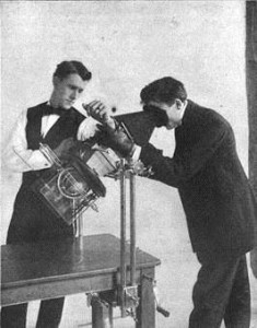 The history of X-rays goes back a long way: they were accidentally discovered by Wilhelm Röntgen in 1895. As it wasn’t clear what the nature of the new radiation was, Röntgen just labelled it ‘X’. The name was supposed to be temporary… but it stuck. As X-rays penetrate soft tissues but not bones, the potential for medical imaging was immediately clear, so much that X-rays were already in use during World War I. Röntgen received the Nobel Prize in Physics for this discovery − and not just a Nobel Prize, but the very first one − in 1901.
An interesting fact is that in the early days there was no suspicion that the new radiation might be harmful, and, for a while, a proper X-rays mania arose, with products advertised as ‘X-rays headache tablets’ and ‘X-rays stove polish’ (and it’s still a mystery what X-rays actually had to do with them). Until the 1950s, X-rays machines could be found in shoe stores, with the purpose of ensuring a perfect fit.
As it soon became clear, X-rays have enormous potential beyond medical imaging as means to shed light on the structure of matter: for example, X-ray crystallography unlocked the structure of DNA in 1953, which led to a Nobel Prize in 1962 (yes, another one. 15 Nobel prizes were awarded for research related to X-rays over the years). The brightest X-rays are found in synchrotron facilities, and the fun fact here is that originally X-rays were an unwanted by-product of particle accelerators developed for high-energy physics (particles moving along a curved trajectory emit radiation, and when their speed is close to that of light this radiation comes in the form of X-rays). Starting from the 1980s synchrotrons entirely dedicated to X-rays came on-line, starting a very productive era of discoveries in materials science, chemistry, biology and drug discovery. Nowadays the first X-ray free electron lasers, which offer ultrafast and very bright X-rays, make it possible to film chemical reactions and determine the structure of single biomolecules.
But the range of X-ray applications does not end here. They are used to investigate the Universe, studying black holes and dark matter, to unveil underpaintings and changes in works of art, revealing what went through an artist’s mind while working on a masterpiece. X-rays allowed us to study Egyptian mummies without unwrapping or damaging them (examples here and here), to read ancient scrolls without unrolling them (here and here), to find out that a wooden statue contained an ancient Chinese mummy… and even to see inside the remains of dinosaurs. And not to mention applications in daily life, such as in airport security.
We went a long way from the very first X-ray image — the hand of Röntgen’s wife — and in directions he would never have imagined.


Please sign in or register for FREE
If you are a registered user on Research Communities by Springer Nature, please sign in