Non-invasive biomarkers and tests are urgently needed for early detection of cancer when it is most treatable. Despite tremendous advance in cancer medicine, unfortunately, very few blood biomarkers have been clinically validated for pre-symptomatic detection of cancer. Many failed studies left people with many questions: Is it because we looked at the wrong biology or simply, we just could not detect nor reliably quantify extremely low-level analytes derived from tiny tumors? Can blood-borne markers allow us to detect early tumors with clinically beneficial diagnostic power and lead-time? If so, what analytical capabilities will be required for quantitative and reliable measurement of these markers?
In 2011, I read a paper which was trying to answer some of these basic questions with a mathematical model of the kinetics of biomarker release by tumors (1). Their model estimated that current clinical techniques needs to be 10,000-fold more sensitive in order to detect blood markers, e.g., CA125 for ovarian cancer, shed from early-stage tumors of submillimeter sizes. Such striking finding was a big motivation that convinced me to develop my independent research dedicated to developing sensitive and quantitative technologies and blood biomarkers to track tumor dynamics for clinical diagnosis and treatment.
A central theme of my research is to explore circulating exosomes as a new dimension of liquid biopsy of tumor, as they provide a unique source of enriched proteins, RNAs and other markers from parental cells. My initial interest in this field arose from a failed experiment that I did during my postdoc training. In 2010, I had developed an ultrasensitive digital PCR technology for single cell mutation analysis. A graduate student from our collaborator’s lab asked me to try her purified extracellular vesicle samples because she had hard time detecting the target fusion gene with standard PCR. I was very confident about the sensitivity of our technology; but all my trials came out negative. Although I didn’t get chance to pursue further in this direction, I was deeply intrigued by the failed experiments and became really interested in these mysterious vesicles that people knew very little about by then.
Eight years later, a work that we published with Dr. Andrew Godwin, who is an oncologist at the University of Kansas Cancer Center, seems to reveal the mystery. When quantifying fusion transcripts in EVs derived by Ewing Sarcoma cells, we found that the level of the transcript is extremely low in EVs, on the order of 10-5 copies per vesicle (2). Based on this result, a quick napkin calculation suggested that my failed tests are likely because I simply didn’t add enough amount of samples to my assays!
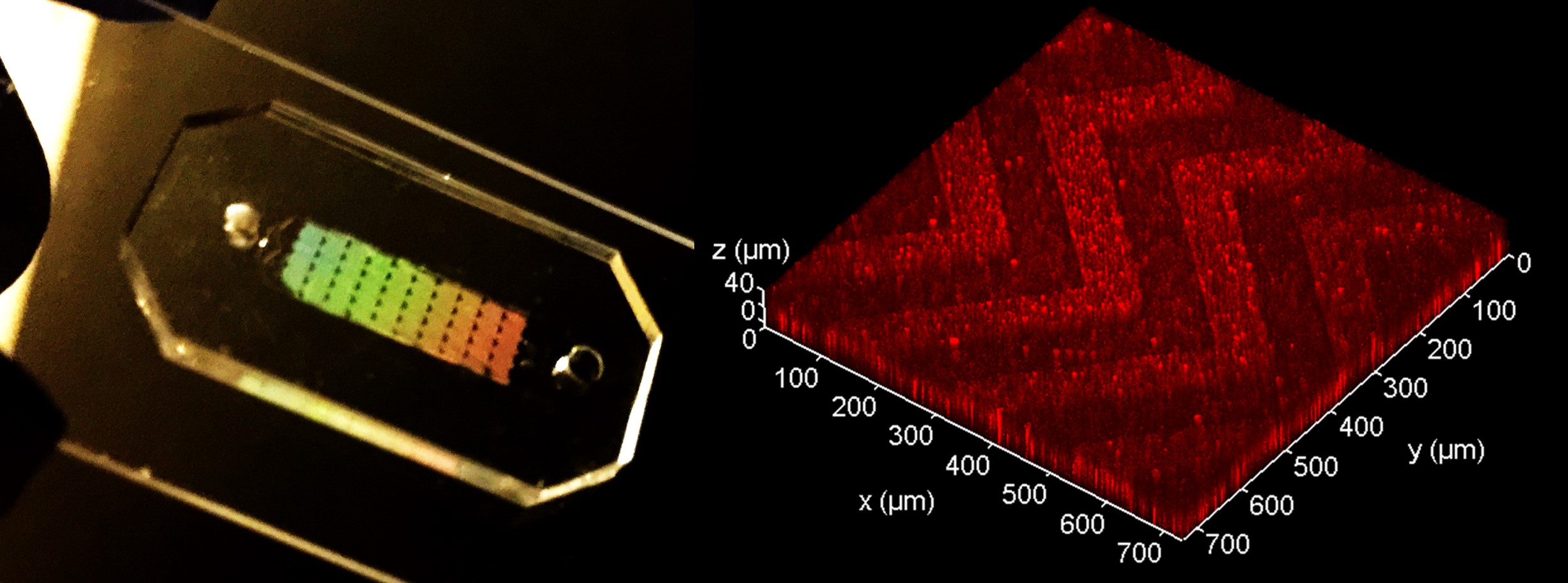
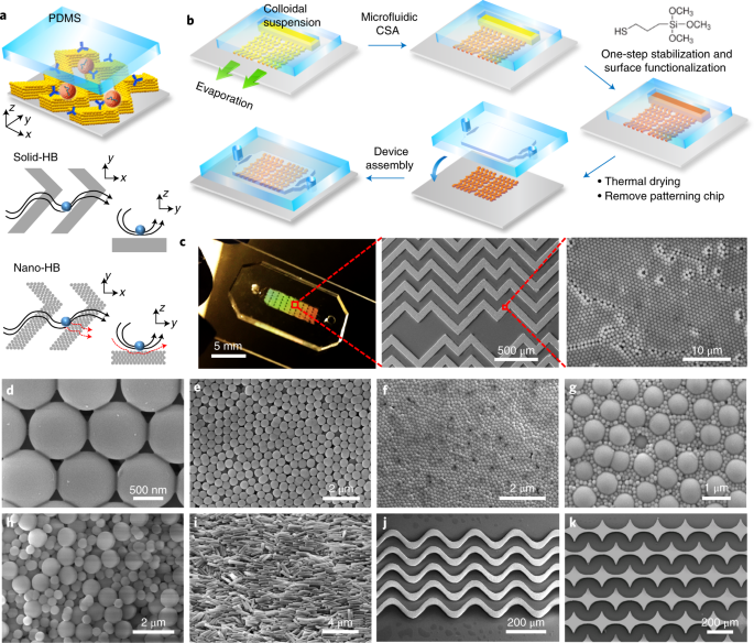
This is one of the examples that manifests the necessity of developing ultrasensitive exosome analysis technologies. The nano-HB chip published in Nature Biomedical Engineering is the third-generation microdevice that we developed for ultrasensitive analysis of circulating exosomes. Inspired by the nanomaterial self-assembly, I came up with an idea to develop an innovative 3D nano-engineering strategy that could yield a unique HB device to enhance exosome analysis by simultaneously overcoming the fundamental limits in mass transfer, surface reaction, and boundary effects. This idea was turned into reality by the joint efforts from all co-authors, especially the first author, Dr. Peng Zhang. Dr. Zhang is an outstanding experimentalist and the best postdoc scholar I have ever worked with. It was his two-month tireless work that overcame the technical bottleneck to integrate and stabilize the self-assembled nanostructures on microfluidic chips. He has spent numerous efforts on improving the device to achieve an impressive sensitivity to detect as low as 10 exosomes per μL sample.
We continue to perfect the chip manufacturing and design and to explore broader applications of the technology via collaborating with biologists and clinicians. Our long-term goal is to develop innovative liquid biopsy tools for disease diagnosis, prognosis and therapy assessment, and translate them into clinical research and treatment strategies.
Our paper: Zhang, P. et al. Ultrasensitive detection of circulating exosomes with a 3D-nanopatterned microfluidic chip. Nature Biomedical Engineering (2019) DOI: https://doi.org/10.1038/s41551-019-0356-9.
1. Hori SS, Gambhir SS. Mathematical Model Identifies Blood Biomarker–Based Early Cancer Detection Strategies and Limitations. Science Translational Medicine. 2011;3(109):109ra16-ra16. doi: 10.1126/scitranslmed.3003110.
2. P. Zhang, J. Crow, D. Lella, X. Zhou, G. Samuel, A.K. Godwin*, Y. Zeng*, “Ultrasensitive Quantification of Tumor mRNAs in Extracellular Vesicles with Integrated Microfluidic Digital Analysis Chip”, Lab on a Chip, 2018, 18, 3790–3801. DOI: 10.1039/C8LC01071D
Follow the Topic
-
Nature Biomedical Engineering
This journal aspires to become the most prominent publishing venue in biomedical engineering by bringing together the most important advances in the discipline, enhancing their visibility, and providing overviews of the state of the art in each field.

Related Collections
With Collections, you can get published faster and increase your visibility.
Implantable wireless communication technologies
Publishing Model: Hybrid
Deadline: Nov 28, 2026
Medical Ultrasound: Emerging Techniques and Applications
Publishing Model: Hybrid
Deadline: Jan 29, 2027


Please sign in or register for FREE
If you are a registered user on Research Communities by Springer Nature, please sign in