Alveolar Ridge Preservation: From Searching for the Best Tissue-Based Approach to Understanding the Evidence

Published in Surgery
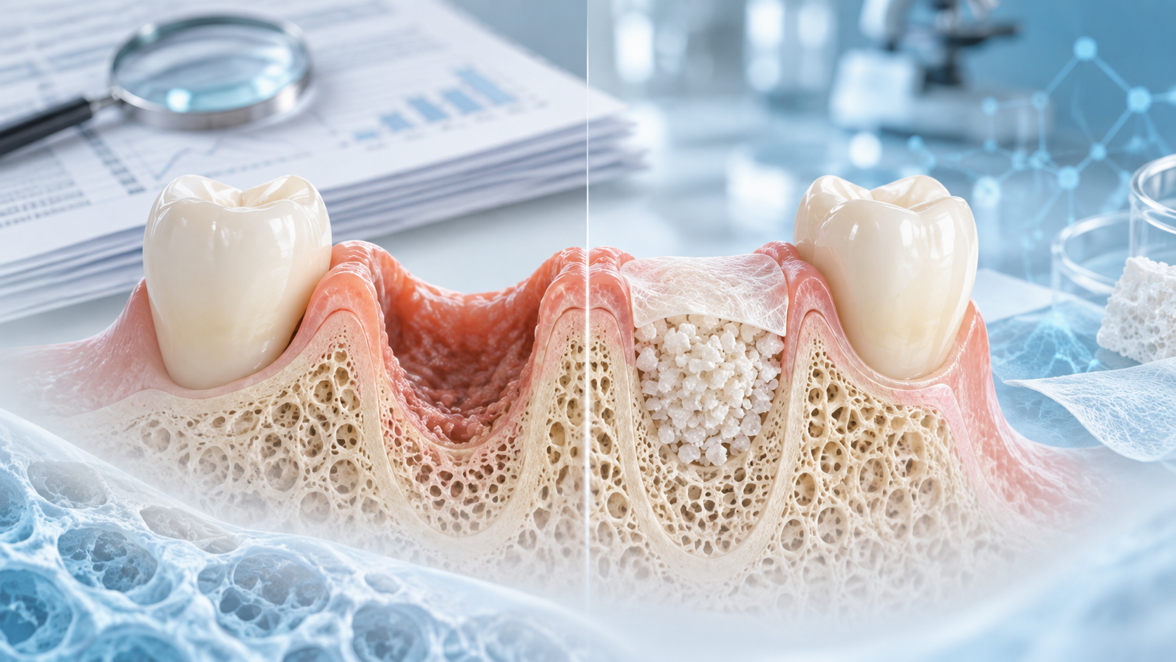
Alveolar Ridge Preservation: From Searching for the Best Tissue-Based Approach to Understanding the Evidence

Share this post

Choose a social network to share with, or copy the URL to share elsewhere

This is a representation of how your post may appear on social media. The actual post will vary between social networks

Explore the Research

Springer Berlin Heidelberg
Springer Berlin Heidelberg Springer Berlin Heidelberg

Alveolar ridge preservation versus unassisted socket healing following tooth extraction: a systematic review of controlled clinical studies and meta-analysis of randomised trials

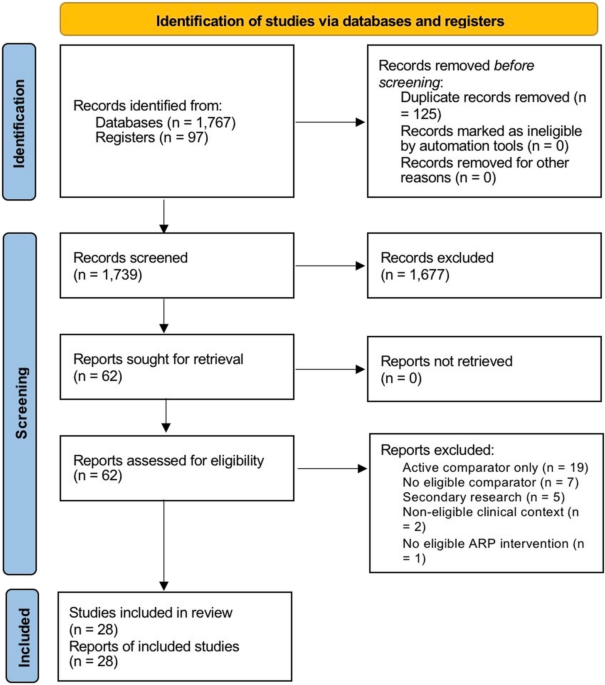
Purpose To estimate early horizontal ridge-width and explicit buccal/facial vertical hard-tissue loss after alveolar ridge preservation (ARP) versus unassisted socket healing. Methods The review was registered in PROSPERO (CRD420261432689). PubMed/MEDLINE, Scopus, Web of Science Core Collection, Embase.com, and ClinicalTrials.gov were searched on 24 June 2026; CENTRAL provided supplementary verification on 1 July 2026. Two reviewers independently selected reports. One reviewer extracted data; the second verified classifications and effect inputs. Randomised direct comparisons were pooled using REML random-effects models with Hartung–Knapp inference. Risk of bias and certainty were assessed with RoB 2 and GRADE. Results Of 1,864 records, 1,739 were screened, and 62 underwent full-text assessment. 28 direct comparison reports were eligible; 20 randomised studies contributed quantitatively, and 8 reports were narrative-only. ARP was associated with less horizontal ridge-width loss (k = 18; MD − 1.37 mm, 95% CI − 1.80 to − 0.94; I² = 75.9%; prediction interval − 2.86 to 0.12) and less explicit buccal/facial vertical loss (k = 13; MD − 1.18 mm, 95% CI − 1.61 to − 0.75; I² = 74.3%; prediction interval − 2.54 to 0.17). Certainty was low for both. The exploratory broad vertical/buccal synthesis favoured ARP (k = 16; MD − 1.26 mm, 95% CI − 1.63 to − 0.89) with very low certainty. Conclusion ARP may reduce early dimensional loss, but substantial heterogeneity and prediction intervals including no effect limit generalisability. Evidence does not establish universal indications, intervention superiority, or long-term implant, aesthetic, or patient-reported benefit.

This review began with a very practical question. I wanted to understand whether one tissue-based approach for alveolar ridge preservation could be considered better than the others after tooth extraction.

At the beginning, my interest was focused on the different regenerative options used to preserve the extraction socket, particularly tissue-based and biologically oriented approaches. I expected that the literature might allow a relatively straightforward comparison between these strategies.

But the deeper I went into the evidence, the clearer it became that the question was more complicated.

The available studies used a wide range of interventions, including collagen matrices, platelet concentrates, biologically active adjuncts, membranes, grafting materials, and combinations of these approaches. They also differed substantially in socket anatomy, healing protocols, follow-up times, and—most importantly—the outcomes they measured. Some studies assessed horizontal ridge loss, others facial or buccal vertical changes, while others focused on histology, mineralised tissue, or residual biomaterial. These outcomes are related, but they are not interchangeable. 

At that point, the purpose of the review evolved.

Rather than trying to identify a “best” material or tissue-based technique from evidence that was too heterogeneous to support such a conclusion, we asked a more fundamental question: what is the direct effect of performing alveolar ridge preservation compared with allowing the extraction socket to heal without an ARP intervention?

This led us to focus on direct ARP-versus-unassisted-healing comparisons, to separate anatomically explicit buccal/facial vertical loss from broader vertical measurements, and to retain relevant evidence narratively when it could not be pooled without unsupported assumptions. 

In the end, the review did not identify a single superior tissue-based approach—and that became an important finding in itself. Instead, it helped clarify what the current evidence supports, where uncertainty remains, and why future research needs more standardised outcomes and better head-to-head comparisons if we truly want to know which regenerative strategy performs best.

For me, this paper became less about finding one “winner” and more about understanding the limits of the evidence—and building a clearer foundation for the next question.

Follow the Topic

Oral and Maxillofacial Surgery
Life Sciences > Health Sciences > Surgery > Oral and Maxillofacial Surgery

Related Collections

With Collections, you can get published faster and increase your visibility.

Current State and Future Opportunities for 3D Printing/Additive Manufacturing in Oral and Maxillofacial Surgery: from Basic Science to Clinical Applications

Three-dimensional (3D) printing, also known as additive manufacturing, has emerged as a transformative technology in oral and maxillofacial surgery. It offers unprecedented precision and customization in the fabrication of patient-specific anatomical models, surgical guides, and implants. This not only enhances preoperative planning and intraoperative accuracy but also significantly reduces surgical time and improves postoperative outcomes, as surgeons can now visualize complex craniofacial structures in tangible form, simulate procedures, and tailor interventions to the unique anatomical and functional needs of each patient. It minimizes risks and optimizes aesthetic and functional results, especially in cases involving congenital deformities, trauma reconstruction, oncologic resections, and orthognathic surgery, where traditional methods often fall short in achieving ideal fit and alignment. The integration of biocompatible materials and advances in digital imaging and computer-aided design (CAD) further expand the scope of 3D printing applications, enabling the production of implants that conform precisely to bone contours and support tissue regeneration. As this technology continues to evolve, with ongoing research into bio-printing and the use of stem cells and growth factors, the potential for creating living tissue constructs and fully functional bone grafts becomes increasingly feasible, promising a future where personalized, regenerative solutions become standard practice in maxillofacial care. Although challenges remain in terms of regulatory approval, cost-effectiveness, and widespread clinical adoption, the growing body of evidence supporting its efficacy and safety underscores the importance of continually educating practitioners on the latest developments in the subject to enable informed decision-making and ultimately, improved patient outcomes.

Publishing Model: Hybrid

Deadline: Ongoing