Design of protein-based stimulus-responsive fractal assemblies
Published in Chemistry

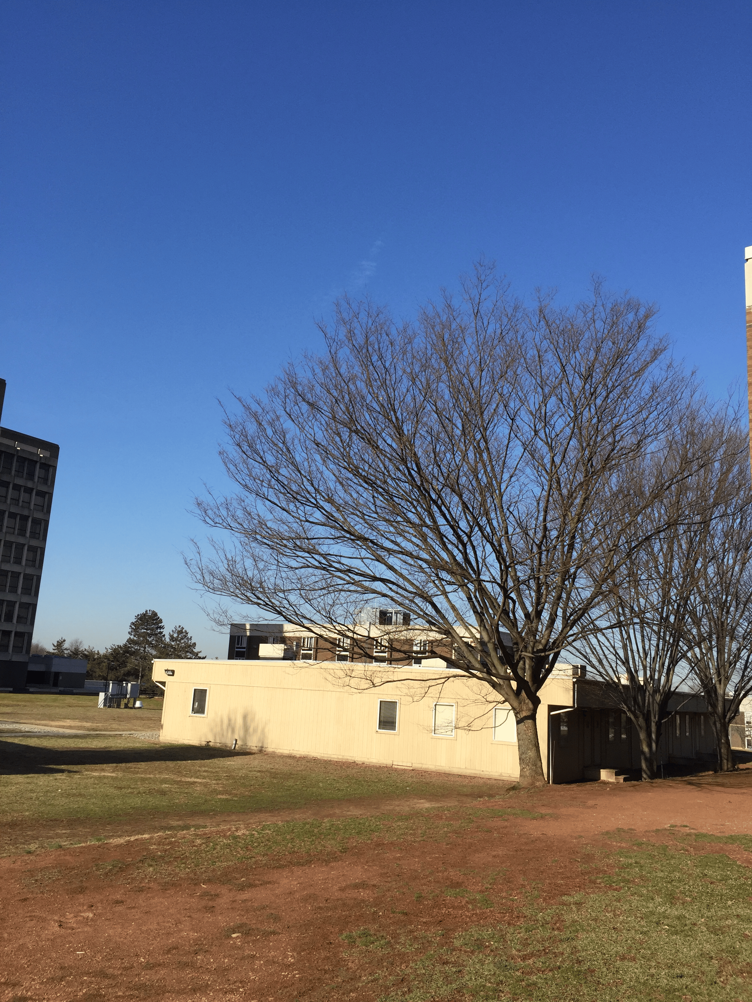
Trees outside a Rutgers building (Photo taken by Sagar Khare, ca 2015)
Some years ago, I moved from the evergreen state of Washington to the North Eastern part of the US to start my lab at Rutgers University. I quickly realized that as much as I enjoyed being a Principal Investigator (PI) and thinking about science 24/7, I needed to have a few hours every week where I could consciously decouple from everything related to my PI-ness. I took to taking walks on Busch campus and in the parks around Rutgers. As the seasons changed (and they really did, unlike in the Pacific North West), the trees transformed from being dense and green to leafless and bare, revealing intricate skeletons that were simultaneously diverse and topologically unique. The shape of the trees became even more stark and beautiful when, during blizzards, a layer of ice highlighted their structure. I couldn’t help but wonder: why are trees shaped the way they are? Is there any advantage to their shape? Trees have another striking feature. A smaller part of the tree often looks like a scaled down version of the entire tree. What causes this unique topological property of trees? The answer, as I discovered upon reading the literature, is that trees are fractional dimensional (fractal) objects which allows them to maximize surface:volume ratios for efficient exchange of matter and energy capture.
Fractal shapes are, in fact, pervasive in geology and in many macroscopic biological objects, including human lungs (which have a surface area the size of a tennis court-- 100 m^2-- in a volume of 6 litres!), and retina. Fractals have remarkable mathematical properties, and their self-similarity has spurred many an artist’s interests.
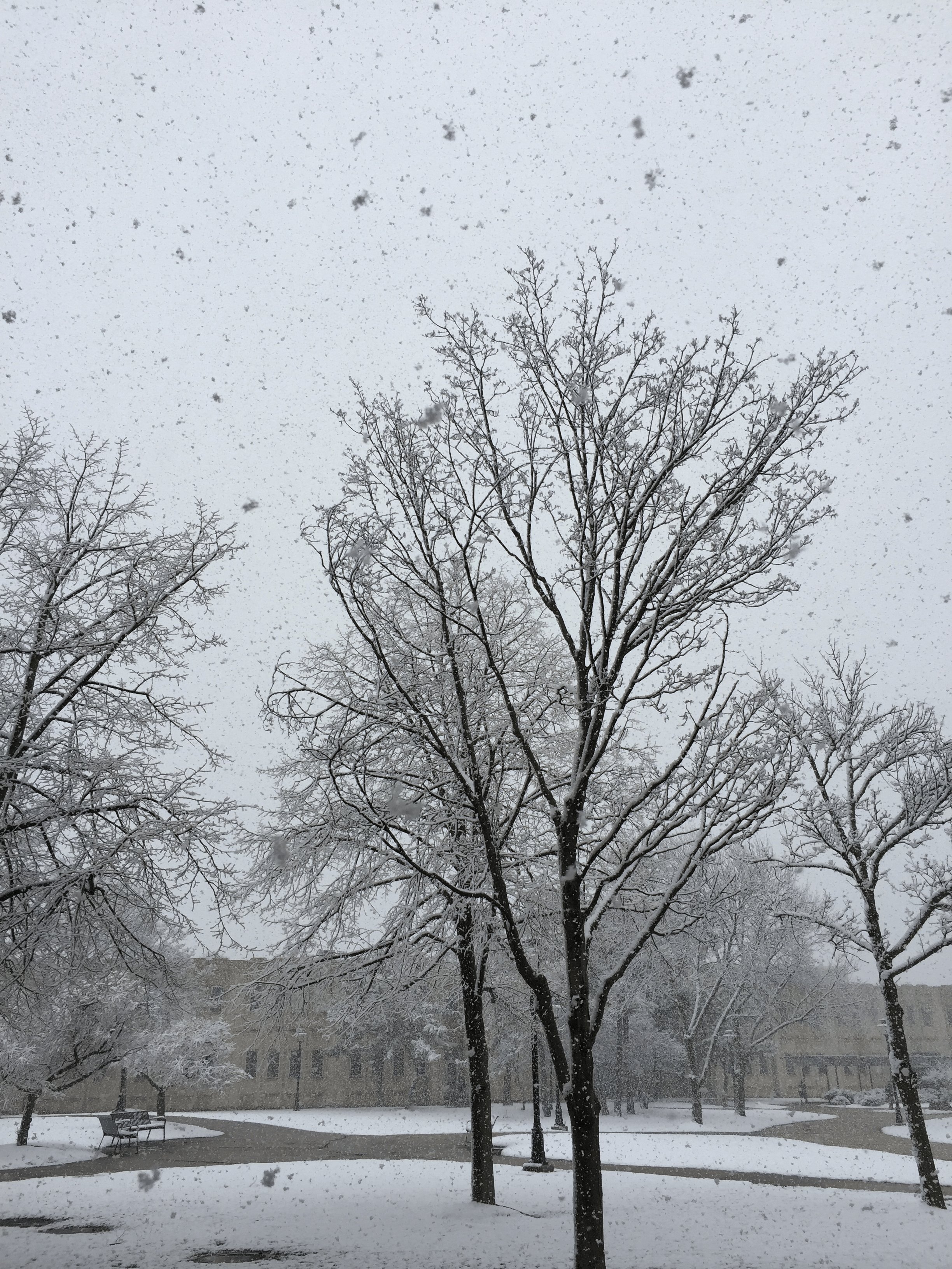
Trees in a snowstorm (Photo taken by Sagar Khare ca 2015)
In a casual discussion (circa 2014) with a friend who had seen a documentary on fractals, the question arose about how fractal shapes are formed, or arise in nature. I had first learnt about fractals in graduate school in the polymer physics class taught by Michael Rubenstein at UNC, and had read more about scaling ideas in graphs in the work of my PhD advisor Nikolay Dokholyan. Nikolay had then pointed me to papers written in the 80s about the formation of fractals (ice flakes, etc.) in the work of my academic grandfather, Gene Stanley. I explained to my acquaintance about some possible models that lead to fractal shapes (e.g., diffusion-limited aggregation model of Witten and Sander), how trees are fractals, and how their shape is functionally useful. “Can you make fractals with proteins, then?” the acquaintance asked. I thought it was an interesting idea, and found myself considering this challenge periodically, especially on my walks over the next few months. If we could make really high surface:volume protein assemblies, that would have many applications as molecular sponges, and for efficient energy capture. But really, wouldn’t it be cool to make beautiful fractal shapes similar to the trees that had long fascinated me? As a (protein) designer, aesthetics were as much a motivating force for me as the potential usefulness of any envisioned assemblies. I filed this idea away as a risky one. “One day, when I have the luxury of tenure…” I thought.
In the Spring of 2015, graduate student Nancy Hernandez and undergraduate student Sophia Tan set up a team representing Rutgers University in the annual BioMolecular Design (BIOMOD) competition held in October. Undergraduate teams spend a summer in a mentor's laboratory trying to build devices made from biomolecules, and then present their work at the BIOMOD Jamboree in October. I thought this could be the perfect opportunity to explore protein assembly formation, and maybe eventually fractals. A few years later (and with tenure, thankfully), due largely to the supremely talented students I was lucky to work with, we were able to realize this vision of computationally designing fractal-shaped assemblies using protein building blocks! There’s much more to do and explore, but I am glad we took the risk.
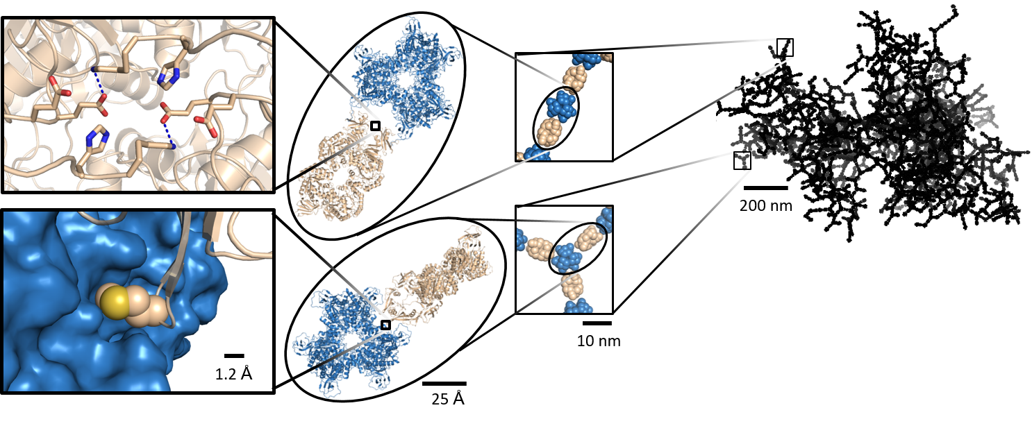
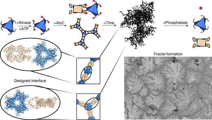
Multi-scale computational design approach for fractal formation
Over to what the students remember:
Denzel Zhu says:
In 2015, the Rutgers BIOMOD Team (led by Sophia Tan; composed of Jason Lin, Maria Shea, Barry Li, Alejandro Herrera, Chris Herrera, and Denzel Zhu) wanted to build protein assemblies using florescent proteins. The idea was that linking together member enzymes of a metabolic pathway into a larger assembly would enable us to increase the efficiency of that reaction pathway. We wanted to prototype our approach with stable florescent proteins (GFP and RFP). The Khare group eventually successfully used this strategy with enzymes for the degradation of the pollutant trichloropropane (TCP).
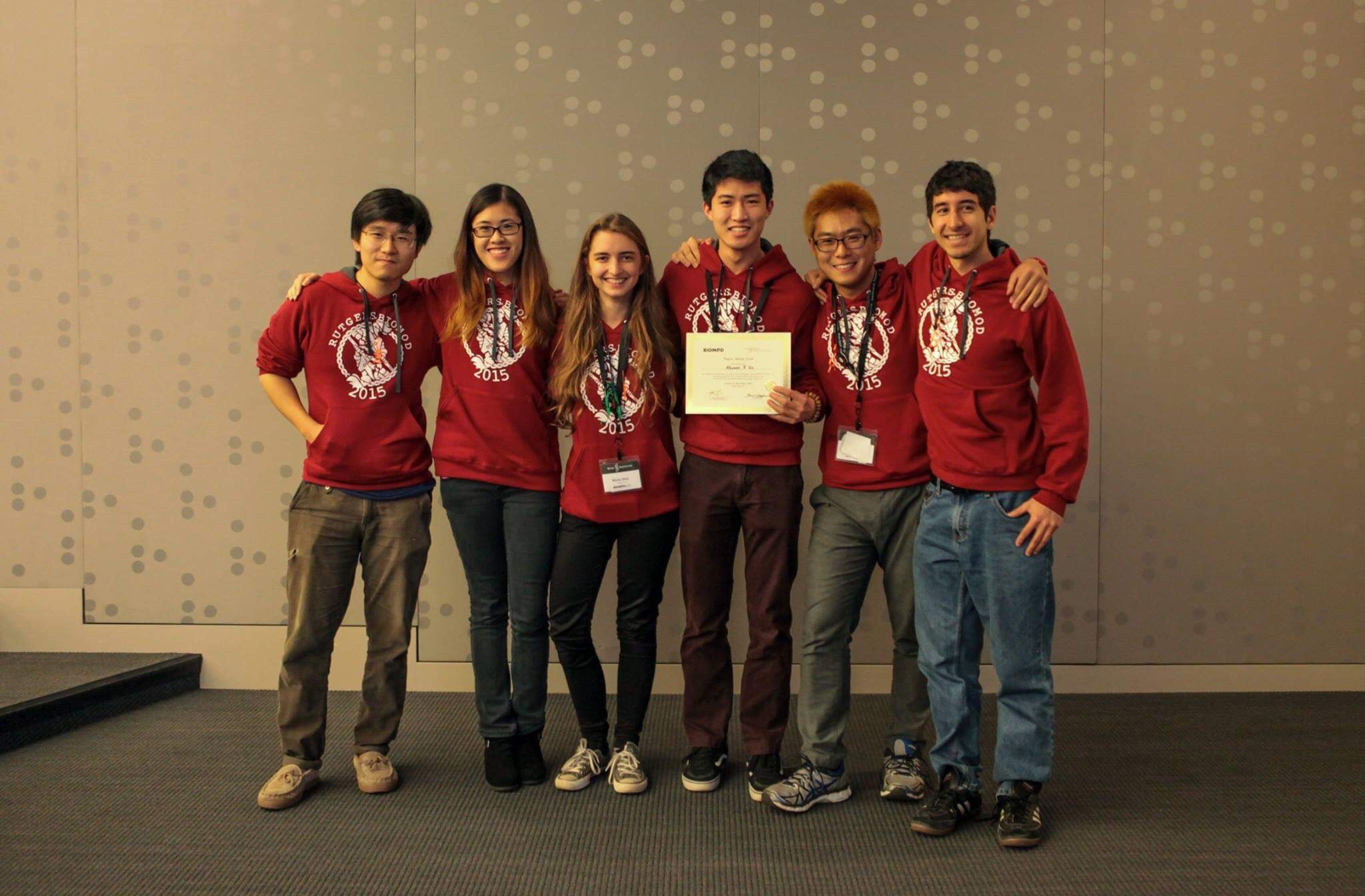
Rutgers BIOMOD Team from 2015
The idea was to use binding domains that had an unnatural amino acid which could form covalent bonds with a binding peptide, and to fuse the binding domain and peptide onto GFP and RFP in order to make oligomers. The binding domain that contained the unnatural amino acid needed to be designed from existing binding domains using software (Rosetta). We successfully produced a binding domain – binding peptide combination that could form covalent interactions, but we found that this approach was impractical for further optimization of assembly formation as the expression yields of unnatural amino acid-containing proteins are too low.
In 2015, we also discovered that divalent connections between component proteins resulted in stable protein assemblies, without the need to form covalent bonds using an unnatural amino acid. With this information in mind, we decided to change our approach to designing a protein assembly: we aimed to maximize divalent connections between oligomeric components. This strategy led to our design of a hexamer – tetramer protein assembly in 2016. We chose AtzA and AtzC as assembly components due to their geometry (hexamer and tetramer, enabling 3 divalent connections between each protein) and because they belonged to the same enzymatic pathway (the degradation of the herbicide atrazine into cyanuric acid).
In 2016, the Rutgers BIOMOD Team (led by Maria Shea and Denzel Zhu; composed of Marium Khalid, Milton Liu, Alisa Permaul, Olivia Dineen, and Grant Bilker) worked on developing a protein assembly with AtzA and AtzC as components. We used protein design software (Rosetta) to devise different linker sequences (the protein sequences used to link binding domains-binding peptides to AtzC/AtzA respectively) to encourage ‘fractal-like’ assemblies, and expressed those engineered proteins. We presented some promising preliminary data at the 2016 BIOMOD Jamboree, but our work on fully experimentally characterizing and optimizing the protein assembly took approximately two more years.
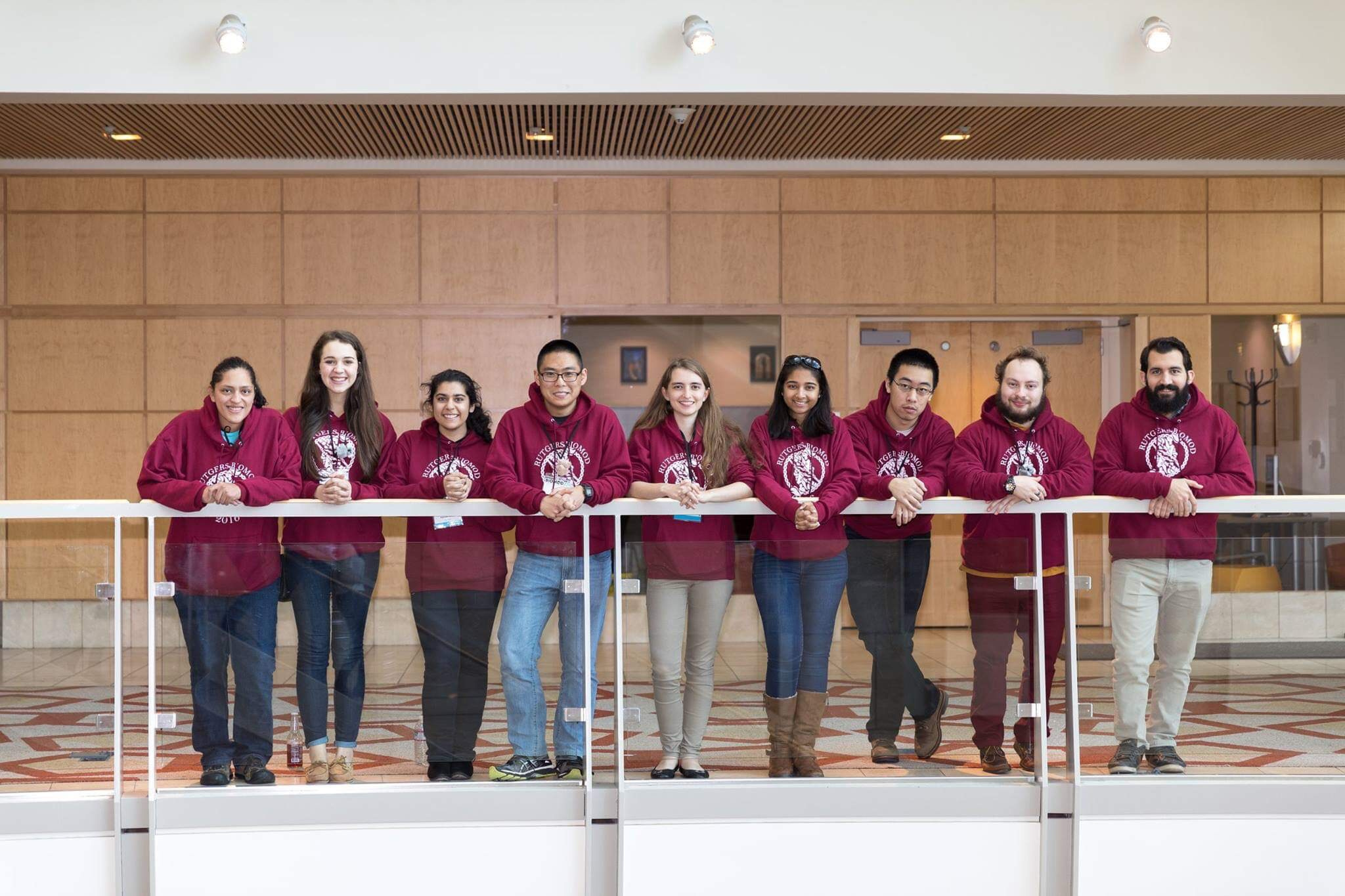
Rutgers BIOMOD team from 2016 with mentors Nancy Hernandez (extreme left) and Will Hansen (extreme right)
Will Hansen says:
In early 2016, Sagar approached me about adapting a program I wrote for making symmetric protein assemblies. The program was intended to insert metal binding sites at the interface of symmetric proteins, much like fitting engine blocks into different car frames, but the underlying symmetry code could be repurposed for the task of designing assemblies. I remember sitting in his office, as we usually do when starting new projects, taking turns drawing on the white board behind his chair, and later using ball-and-stick models to argue about which degrees of freedom are redundant for the design of assemblies (see below). It was in one of these initial meetings that we started using our symmetry/oligomer "gang signs". If you ever happen to catch us talking and we have our fingers outs like a pair of battling scissors, rest assured -- hopefully -- good science is underway!
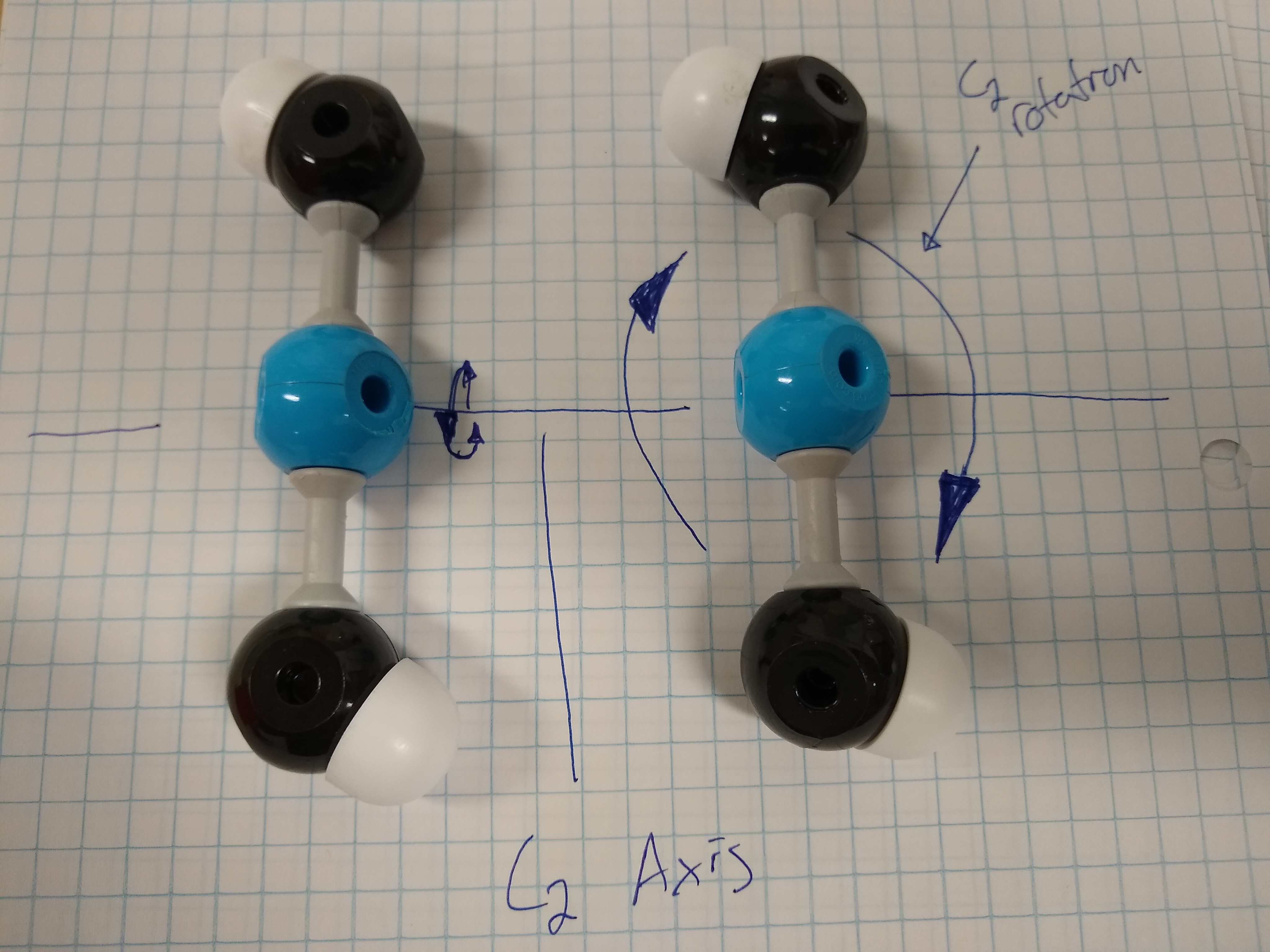
Kendrew models used to visualize relevant degrees of freedom in design
Nancy Hernandez says:
We needed to develop methods to generate fractal enzyme assemblies in order to take advantage of the functional advantage of such a geometry. But, first, we needed to really understand fractals and what it meant to be “fractal-like”. Digging into the literature, we quickly realized that designing a fractal-like structure had never been done with protein building blocks. However, we hypothesized that it should be possible to build these structures since there were natural proteins that formed fractals under some conditions (e.g., silicatein and the silk protein sericin). With help from collaborators, I performed a lot of experimental work to confirm fractal formation, and to demonstrate control over formation of these assemblies. I was particularly excited when we saw the same structures using multiple microscopy techniques, and when our simulations agreed with data from cryo-electron tomography. I am looking forward to seeing how the lab takes our studies forward and (hopefully) builds fractal topologies inside cells one day!
Follow the Topic
-
Nature Chemistry
A monthly journal dedicated to publishing high-quality papers that describe the most significant and cutting-edge research in all areas of chemistry, reflecting the traditional core subjects of analytical, inorganic, organic and physical chemistry.


Please sign in or register for FREE
If you are a registered user on Research Communities by Springer Nature, please sign in