Fascinating scintillation performances in lanthanide doped fluoride nanoparticles
Published in Materials

X-rays are electromagnetic waves with short wavelengths and strong penetrability in physical matter, including live organisms. Scintillators capable of converting X-rays into the ultraviolet (UV), visible or near-infrared (NIR) photons are widely employed to realize indirect X-ray detection and XEOL imaging in many fields. They include medical diagnosis, computed tomography (CT), space exploration, and non-destructive industrial material and security inspections.
Commercial bulk scintillators possess high light yield (LY) and superior energy resolution. However, they suffer from serval drawbacks, i.e., complex fabrication procedures, expensive experimental equipment, non-tunable XEOL wavelength and poor device processability. They all produce emissions in the visible spectral range, but having XEOL in the NIR range may find more interesting applications in biomedicine. Thick crystals also generate light scattering followed by evident signal crosstalk in a photodiode array. Recently, metal halide perovskites have been investigated for X-ray detection. Unfortunately, these materials also exhibited some intrinsic limitations, such as poor photo-/environmental- stability, heavy metal toxicity and low LY. Thus, the search for developing a new generation of scintillators is still a considerable focus of scientific research.
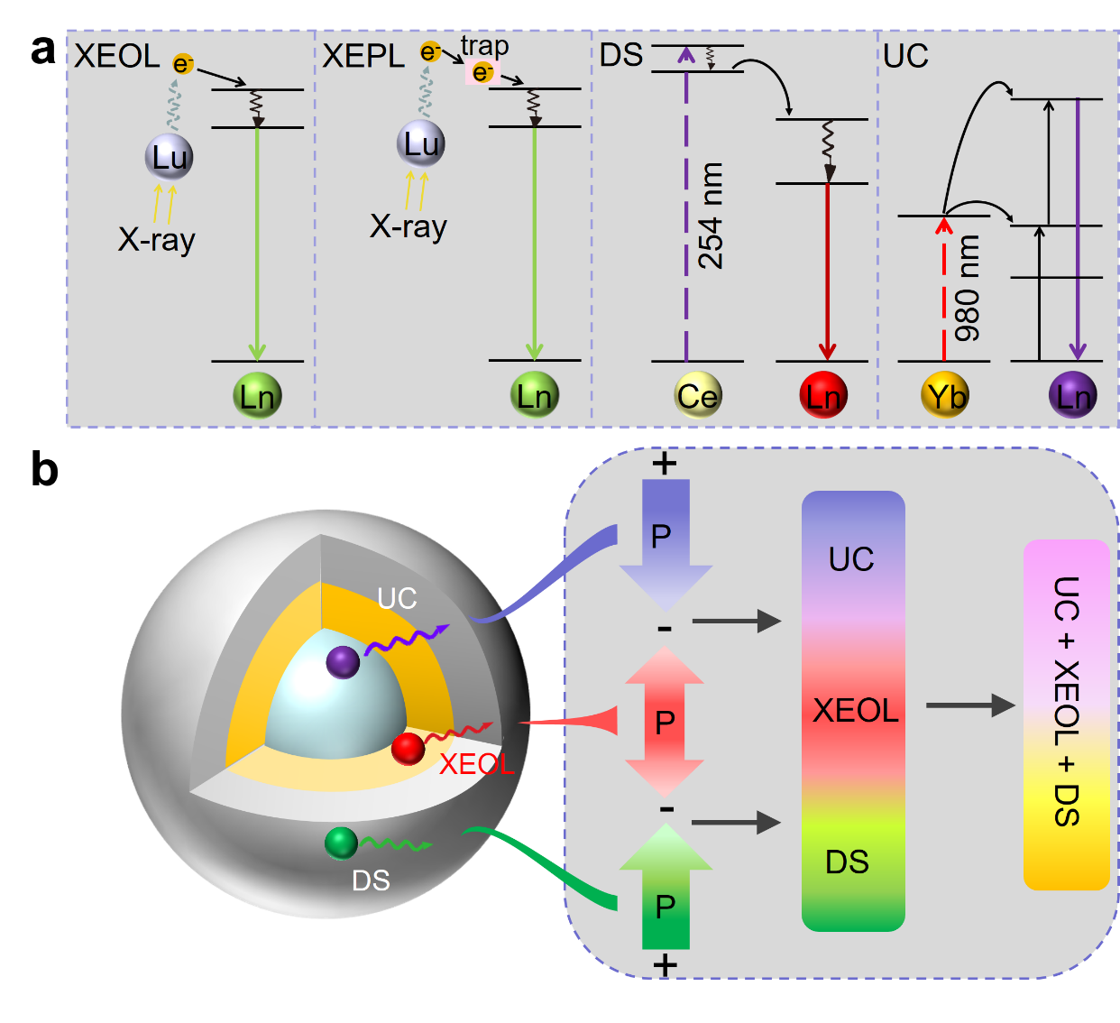
Lanthanide-doped fluoride NSs avoid the limitations of bulk scintillators and metal halide perovskites. They also exhibit many useful properties. The core-shell structures of the lanthanide doped fluoride NSs can be tuned and designed on demand by employing a cheap and convenient wet-chemical method. The emission wavelengths can be tuned and extended to the second NIR window, benefiting from the abundant energy levels of lanthanide activators (Fig. 1). These NSs show superior photostability, low toxicity and convenient device processability. It makes them promising candidates for next-generation NSs and XEOL imaging. Moreover, they exhibit XEPL property, showing promising applications in biomedicine and optical information encoding. The combination of XEOL and XEPL makes them suitable for broadening the scope of their applications. This review discusses and summarizes the XEOL and XEPL characteristics of lanthanide-doped fluoride NSs. The review ends with a discussion of the existing challenges for advancing this field.
Lanthanide doped fluoride NSs have been broadly used to generate NIR triggered photon UC and UV excited DS emissions in addition to XEOL and XEPL (Fig. 1a). Core/multi-shells nanostructure can be employed to simultaneously produce XEOL, XEPL, as well as UC and DS, which allow researchers to manipulate the excitation dynamics of lanthanide activators. For example, when different lanthanide activators are used to generate diverse emission wavelengths of XEOL, UC and DS in core@shell@shell NSs (Fig. 1b), plentiful multicolors can be modulated on demand through controlling the excitation wavelength and/or power, which show promising applications in dynamic display and multi-level anti-counterfeiting. Especially, through modulating the incident photons at different timescale based on a specific requirement, time-response characteristic transitions can be designed, which show promising in multi-mode bio-imaging and bio-sensing.

Fig. 1 a XEOL, XEPL, DS and UC processes in lanthanide doped fluoride NSs. b Schematic illustration of the multimode color evolution based on fluoride core@shell@shell NSs. P represents excitation power.
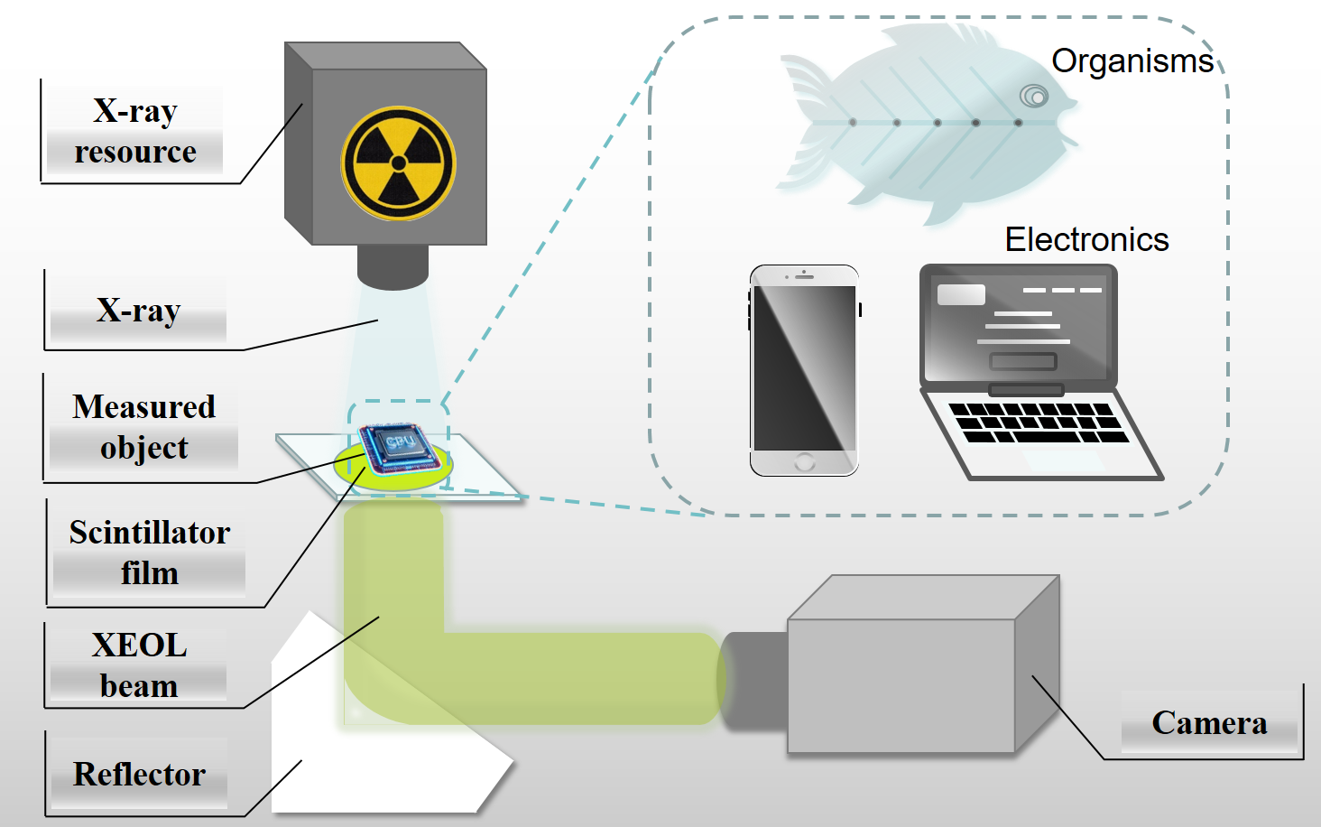
The fundamental working principle of XEOL imaging is to record the attenuation of X-rays after penetrating the specific subjects by using a scintillator and then imaging with a camera (Fig. 2). The scintillator screen is placed under the target to absorb the transmitted X-ray photons. For examples, a low dose of X-rays penetrating live organisms enables the application of computed tomography, while penetrating nonliving matter enables product quality and security inspection. The X-ray irradiation dose should be low enough to assure the safety, while the high resolution and distinct contrast are important for image analysis. The maximum permissible dose for gonads and red bone marrow is ~5 rem/year; for skin, bone and thyroid is ~30 rem/year; for hands, forearms, feet and ankles is ~75 rem/ year; for any other single organ is ~15 rem/year.

To promote the development of high-performance fluoride NSs and their practical applications, the team discussed the existing challenges and future multidisciplinary opportunities in this field below. Understanding the XEOL mechanism benefits the design and exploration of new fluoride NSs. At present, how the generated low kinetic energy charge carriers are transported to the luminescent centers or captured by defects and the corresponding influence factors are unclear. The first populated nonradiative excited levels and the radiative levels of lanthanide activators are optimal when calculating or characterizing the energy differences among these charge carriers. These calculations will guide the design of energy transfer processes to match the energy differences followed by the enhanced light yield. High LY is a prerequisite for the realization of ultra-low dose rate applications.
Moreover, the life-times of 4f-4f transitions in most trivalent lanthanide ions vary from a few µs to tens of ms, which are not suitable for real-time dynamic XEOL imaging. Although the decay rate of Ce3+ ions is in the nanosecond range, XEOL emission in fluoride NSs doped by cerium ions is in the UV region, which do not match well with commonly used visible detectors. So to achieve bright XEOL intensity with fast decay rate and appropriate emission wavelength range, it is better to design the local crystal environment of 5d orbitals of Ce3+ and Eu2+ ions in fluoride NSs for the realization of better XEOL performances.
More discussions were described in our paper in detail. Welcome all authors to discuss and cooperate with us in the filed of lanthanide doped nanoscintillators.
Follow the Topic
-
Light: Science & Applications

A peer-reviewed open access journal publishing highest-quality articles across the full spectrum of optics research. LSA promotes frontier research in all areas of optics and photonics, including basic, applied, scientific and engineering results.


Please sign in or register for FREE
If you are a registered user on Research Communities by Springer Nature, please sign in