From body signals to brain rhythms: vagus nerve stimulation, vascular motion, and learning
Published in Biomedical Research

Explore the Research
S2589004226027914?utm_campaign=related_content&utm_source=HEALTH&utm_medium=communities
What makes the brain ready to learn?
We often think of learning as a property of neuronal circuits: synapses change, activity patterns are modified, and behavior improves. This view is, of course, essential. But in our laboratory, we have become increasingly interested in a complementary question: what is the state of the local brain environment in which plasticity occurs?
Neurons do not work in isolation. Their activity depends on oxygen, glucose, metabolic substrates, local pH, glial support, vascular dynamics, and the broader physiological state of the body. In this sense, learning may not be determined only by which synapses are active, but also by whether the surrounding brain environment is permissive for long-term plasticity.
A body-to-brain pathway into the learning brain
This idea led us to study vagus nerve stimulation, or VNS. The vagus nerve is a major communication pathway between the body and the brain. It carries signals from internal organs and bodily states to the brainstem, and from there to widespread brain regions through neuromodulatory systems. Clinically, VNS is already used for conditions such as drug-resistant epilepsy, but how it changes brain function remains incompletely understood.
In our new iScience paper, we asked whether VNS delivered after learning could influence long-term memory formation, and whether this effect might be accompanied by changes in vascular dynamics in the brain.
Stimulating the vagus nerve after training
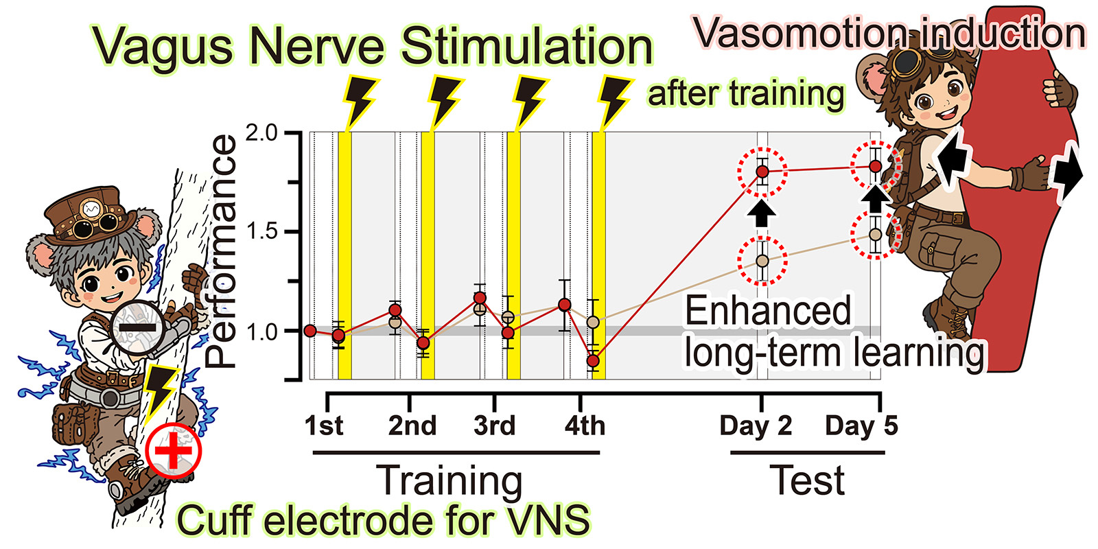
We used a mouse motor learning task called horizontal optokinetic response learning. In this task, mice improve their eye movements in response to moving visual patterns. We delivered VNS immediately after training sessions, rather than during the training itself. This timing was important because we wanted to ask whether VNS affects the processes that occur after learning experience, such as delayed plasticity or memory consolidation.
The result was striking. Post-training VNS enhanced multi-day motor learning. The improvement was not immediate during the training session, but emerged later, across days. This suggested that VNS may influence delayed processes that support long-term learning.
Vascular rhythms in the cerebellum
At the same time, we monitored vascular-related signals near the cerebellar flocculus, a brain region important for this form of motor learning. Using fiber photometry and vascular imaging, we found that individual VNS trains induced transient vascular volume changes. Across repeated stimulation, these responses developed into rhythmic vascular oscillations. Importantly, the magnitude of these vascular oscillations was associated with later learning performance.
What we can—and cannot—conclude
This does not mean that we have proven vascular oscillations cause better learning. We have not. In the paper, we are careful to state that the relationship is correlational. We also did not directly measure ATP, lactate, pyruvate, or oxygen metabolism in this study. What we can say is that VNS enhanced long-term learning and induced structured vascular dynamics, and that these vascular dynamics were linked to later behavioral performance.
For us, this finding is exciting because it suggests that vascular dynamics may be more than a passive consequence of neuronal activity. They may be part of a broader brain-environmental state that accompanies, and perhaps supports, long-term plasticity.
Connecting VNS to REM sleep metabolism
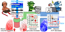
This new work also connects to another recent study from our laboratory on REM sleep metabolism. In that study, published in Communications Biology, we monitored neuronal ATP, astrocytic pyruvate, and cerebral blood volume across sleep-wake states. During REM sleep, cerebral blood volume and astrocytic pyruvate increased, while neuronal ATP decreased. We called this combination an “energy paradox” in REM sleep.
At first glance, the REM sleep study and the VNS learning study may seem different. One is about sleep and brain metabolism; the other is about peripheral nerve stimulation and motor learning. But to us, they are part of the same larger question.
How do brain states, body-derived signals, vascular dynamics, and metabolic support interact to shape brain function?
During REM sleep, the brain appears metabolically active, yet neuronal ATP can fall despite increased blood volume and astrocytic pyruvate. During post-training VNS, body-derived afferent signals induce vascular oscillations and are associated with improved long-term learning. In both cases, neuronal computation is embedded in a changing vascular and metabolic environment.
Toward a brain-environment view of plasticity
We are now extending this line of work by monitoring additional metabolic and vascular signals during VNS, sleep, and learning. Ultimately, we hope to understand whether the brain’s capacity for plasticity depends not only on neuronal firing patterns, but also on the dynamic support provided by glia, blood vessels, and the body.
This study was led by Junyu U. Chen, together with Yoko Ikoma and Ko Matsui, at the Super-network Brain Physiology laboratory, Tohoku University. It reflects our continuing effort to understand the brain not as an isolated organ, but as a dynamic system shaped by signals from the body and by the local environments surrounding neurons.
Signals from the body may echo through the brain’s landscape. By following those echoes in vascular and metabolic rhythms, we may begin to understand how the brain becomes ready to learn.
Paper:
Chen JU, Ikoma Y*, Matsui K* (2026)
Vagal nerve stimulation induces vascular oscillations and enhances long-term learning.
iScience, 117413.
https://doi.org/10.1016/j.isci.2026.117413
Related paper:
Takahashi Y, Ikoma Y, Matsui K* (2026)
Energy paradox in REM sleep: balancing supply and consumption in brain metabolism.
Communications Biology, 9: 979.
https://doi.org/10.1038/s42003-026-10646-6
Join the discussion and share the study on X:
The brain and mind are not shaped by neurons alone. I am Ko Matsui, Professor of Super-network Brain Physiology at Tohoku University (Sendai, Japan). My laboratory studies the mind through whole-body super-networks involving neurons, glia, blood vessels, and peripheral nerves. We welcome graduate students who are eager to explore the frontiers of the science of mind.