From the Clinic to the Molecule: What Movement Disorders Teach Us About Translational Neuroscience
Published in Biomedical Research

One diagnosis, many trajectories
Parkinson’s disease (PD) is the neurological disorder with the fastest-growing prevalence. A modeling study using Global Burden of Disease 2021 data projected that 25.2 million people worldwide would live with PD in 2050 (95% uncertainty interval 21.7–30.1 million), a 112% increase from 2021, driven primarily by population aging (Su et al., 2025). These are model-based projections, not direct observations of future cases.
Behind one diagnostic label lie substantial differences in age at onset, motor phenotype, progression, cognitive course, and response to levodopa.
When two people with the same diagnosis follow different trajectories, a fundamental question arises: do distinct biological processes drive their clinical differences?
Movement disorders provide a useful setting to explore how translational neuroscience can address this question without confusing association, prediction, and causation.
What genetic testing finds and what it does not
Seven genes — LRRK2, GBA1, PRKN, SNCA, PINK1, PARK7 and VPS35 — are widely recognized as established contributors to monogenic forms or genetic risk of PD, although their inheritance patterns and clinical implications differ. In the PD GENEration study, 1,070 of 8,301 participants (12.9%) had a reportable genetic finding. Among 4,406 participants without early onset, an affected first-degree relative, or high-risk ancestry, the yield was still 9.1% (399 participants) (Cook et al., 2024).
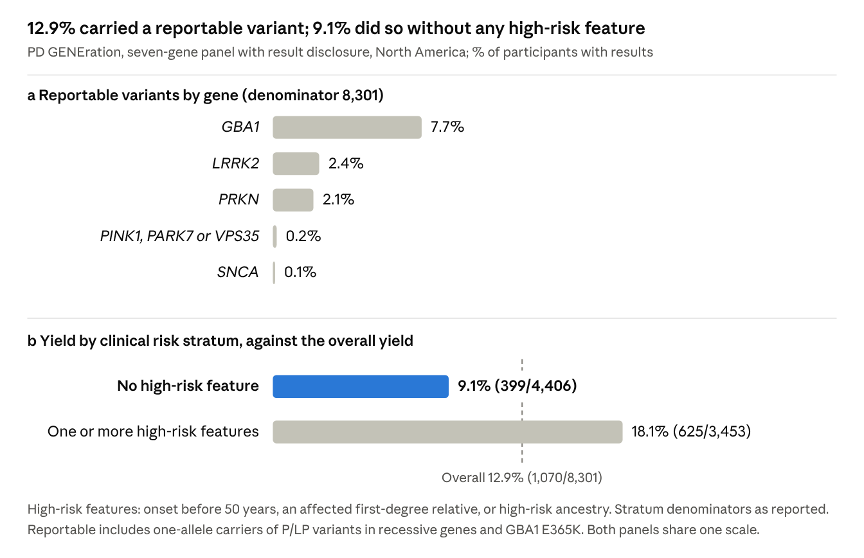 Figure 1 · Genetic findings in PD GENEration. Reportable-result yield in the overall cohort and in participants without specified high-risk characteristics. Data from Cook et al. (2024); original visualization by the author.
These figures require context. The study defined a reportable result as findings that do not necessarily establish a monogenic diagnosis, such as a single variant in a recessive gene or the GBA1 E365 K risk variant. Participants were recruited through clinical sites and self-referral, so these results should not be interpreted as estimates of monogenic PD prevalence in the general population.
Genetic testing can identify a finding that warrants further interpretation; it does not automatically establish the cause of an individual’s disease.
From gene to pathway, and back to the variant
The discovery of PINK1 mutations in families with autosomal recessive early-onset PD helped connect clinical genetics with mitochondrial quality control (Valente et al., 2004). Under mitochondrial stress, PINK1 accumulates on the outer mitochondrial membrane and phosphorylates ubiquitin and Parkin at serine 65. This signaling promotes Parkin activation and amplifies ubiquitin-dependent signals that recruit the autophagy machinery to damaged mitochondria (Koyano et al., 2014; Ordureau et al., 2014; Lazarou et al., 2015).
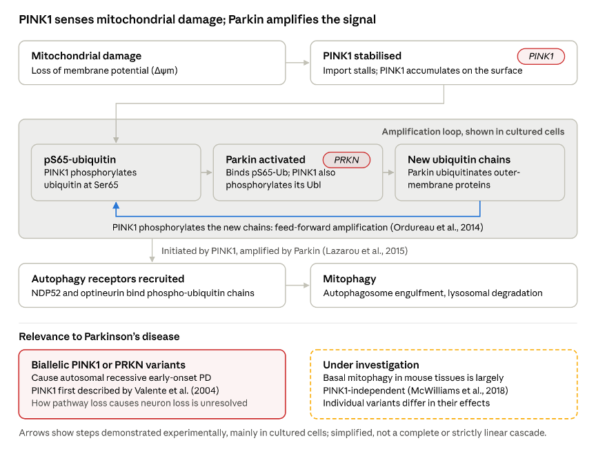 Figure 2 · A simplified schematic of mitochondrial damage signaling, PINK1-dependent phosphorylation, Parkin activation, and recruitment of the autophagy machinery. Based on Koyano et al. (2014), Ordureau et al. (2014) and Lazarou et al. (2015); original schematic by the author.
Experimental evidence strongly supports the pathway, but its operation in living organisms is more complex than a single linear model suggests. For example, basal mitophagy in mouse tissues with high metabolic demand has been observed to proceed largely independently of PINK1 (McWilliams et al., 2018).
Individual variants also differ. A missense change may affect kinase activity, protein stability or mitochondrial import, or have no measurable functional effect. Whether a particular variant disrupts PINK1 signaling or Parkin recruitment is an experimental question, not a conclusion that computational prediction alone can support.
Variant interpretation is evidence integration
The ACMG/AMP framework formalizes how evidence from population frequency, computational prediction, functional studies, segregation, and phenotype can contribute to variant classification (Richards et al., 2015). Evidence is weighted by type, direction, and strength; criteria are combined using defined rules and expert judgment, not simply added as points.
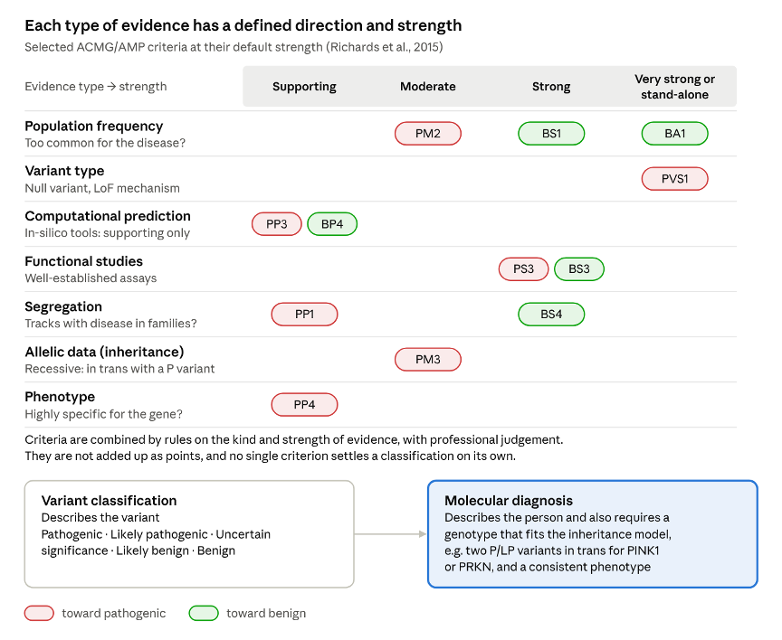 Figure 3 · Integrating evidence in variant interpretation. A schematic showing how population, computational, functional, segregation and phenotype evidence contribute to classification under the ACMG/AMP framework. Based on Richards et al. (2015); original schematic by the author.
One distinction matters: variant classification is not the same as a molecular diagnosis. For recessive PINK1- or PRKN-associated disease, a diagnosis generally requires two disease-associated variants consistent with the inheritance model, usually on opposite alleles. A single heterozygous finding is not sufficient. Public databases such as ClinVar make submitted interpretations and supporting evidence accessible, but differences between submissions underline the need to evaluate the evidence rather than uncritically accept a classification (Landrum et al., 2018).
Measuring what we mean
Freezing of gait (FOG) illustrates how measurement shapes clinical conclusions. A meta-analysis of 29 studies estimated an overall prevalence of 39.9% in PD (95% confidence interval 35.3–44.5%). Estimates varied with the assessment method: 43.8% using item 3 of the Freezing of Gait Questionnaire and 36.0% using item 14 of the Unified Parkinson’s Disease Rating Scale (Ge et al., 2020). These instruments ask different questions, so their differing estimates do not establish that either is superior.
Both items record whether freezing occurs and how often. Wearable inertial sensors could also record when episodes start, how long they last, and where they occur (Figure 4). Across detection studies, reported sensitivities range from 63% to 100%, but samples are small, protocols and algorithms vary, and validation in free-living settings remains limited (Huang et al., 2023).
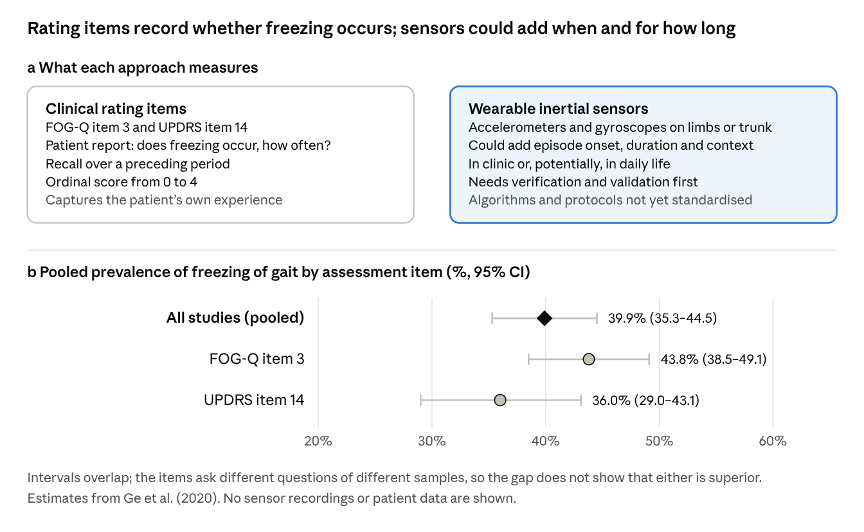 Figure 4 · Measuring freezing of gait. A comparison of clinical rating scales and wearable inertial sensors, illustrating the complementary information they can provide. Prevalence estimates from Ge et al. (2020); the relevant systematic review should support these sensor concepts. Original schematic by the author
Clinical scales capture reported or observed symptoms, whereas wearable sensors can provide continuous information about movement patterns and episode timing. However, technical feasibility is not equivalent to clinical validity. Sensor-derived measures need adequate verification, analytical validation, reproducibility, and clinical validation before they can be relied upon for individual decisions. For FOG, such technologies remain promising tools for research and monitoring, but their performance and usefulness depend on the sensor, algorithm, setting, and intended application (Huang et al., 2023; Goldsack et al., 2020).
Principles for the next question
Across genetics, cell biology, and digital measurement, four principles can help keep translational inference rigorous:
- Match the evidence to the claim. Frequency, prediction, functional impairment, and segregation answer different questions.
- Make the inheritance model explicit. One heterozygous variant in a recessive disease gene is not, by itself, a diagnosis.
- Establish reliability before interpreting differences. A measure that does not reproduce cannot support robust biological conclusions.
- Treat uncertainty as a guide to the next experiment. An uncertain variant or inconclusive functional assay identifies what remains to be tested.
The clinic and the molecule are not opposite ends of a one-way road. Clinical observations generate hypotheses; genetic, experimental, and computational methods test them; and the results inform diagnosis, counseling, and research design. In movement disorders, where clinical diversity may reflect multiple biological processes, progress depends not only on collecting more data but also on making proportionate claims from the evidence. That iterative process is central to translational neuroscience's promise.
References
Cook, L. et al. (2024). Parkinson’s disease variant detection and disclosure: PD GENEration, a North American study. Brain, 147(8), 2668–2679. https://doi.org/10.1093/brain/awae142
Ge, H.-L. et al. (2020). The prevalence of freezing of gait in Parkinson’s disease and in patients with different disease durations and severities. Chinese Neurosurgical Journal, 6, 17. https://doi.org/10.1186/s41016-020-00197-y
Goldsack, J. C. et al. (2020). Verification, analytical validation, and clinical validation (V3): The foundation of determining fit-for-purpose for biometric monitoring technologies. npj Digital Medicine, 3, 55. https://doi.org/10.1038/s41746-020-0260-4
Huang, T., Li, M. and Huang, J. (2023). Recent trends in wearable devices used to detect freezing of gait and falls in people with Parkinson’s disease: A systematic review. Frontiers in Aging Neuroscience, 15, 1119956. https://doi.org/10.3389/fnagi.2023.1119956
Koyano, F. et al. (2014). Ubiquitin is phosphorylated by PINK1 to activate parkin. Nature, 510, 162–166. https://doi.org/10.1038/nature13392
Landrum, M. J. et al. (2018). ClinVar: Improving access to variant interpretations and supporting evidence. Nucleic Acids Research, 46(D1), D1062–D1067. https://doi.org/10.1093/nar/gkx1153
Lazarou, M. et al. (2015). The ubiquitin kinase PINK1 recruits autophagy receptors to induce mitophagy. Nature, 524, 309–314. https://doi.org/10.1038/nature14893
McWilliams, T. G. et al. (2018). Basal mitophagy occurs independently of PINK1 in mouse tissues of high metabolic demand. Cell Metabolism, 27(2), 439–449.e5. https://doi.org/10.1016/j.cmet.2017.12.008
Ordureau, A. et al. (2014). Quantitative proteomics reveal a feedforward mechanism for mitochondrial PARKIN translocation and ubiquitin chain synthesis. Molecular Cell, 56(3), 360–375. https://doi.org/10.1016/j.molcel.2014.09.007
Richards, S. et al. (2015). Standards and guidelines for the interpretation of sequence variants: A joint consensus recommendation of the ACMG and AMP. Genetics in Medicine, 17(5), 405–424. https://doi.org/10.1038/gim.2015.30
Su, D. et al. (2025). Projections for prevalence of Parkinson’s disease and its driving factors in 195 countries and territories to 2050: Modelling study of Global Burden of Disease Study 2021. BMJ, 388, e080952. https://doi.org/10.1136/bmj-2024-080952
Valente, E. M. et al. (2004). Hereditary early-onset Parkinson’s disease caused by mutations in PINK1. Science, 304(5674), 1158–1160. https://doi.org/10.1126/science.1096284
All four figures are original graphics by the author, drawn from the published results and sources cited in each caption; no published figure is reproduced.