From the laboratory to the free-throw line: measuring brain and body in motion
Published in Social Sciences, Neuroscience, and Anatomy & Physiology



Most of what we know about the human brain comes from laboratory experiments in which participants are asked to remain as still as possible. This control is essential for many forms of neuroscience and neuroimaging, but everyday human behavior rarely occurs without movement. Walking, playing sports, and interacting with our surroundings require continuous coordination between the brain and the body.
Our study began with a methodological question: could we record the brain activity that precedes a complex movement in a natural setting outside the laboratory?
As silly as it may sound, the corresponding author thought that basketball free throws would offer a useful test. A free throw is familiar to experienced players, goal-directed, and repeatable, but it still requires a coordinated sequence of postural adjustments and movements of the arms, hands, and legs. Rather than bringing basketball players into an artificial laboratory simulation, we wanted to bring a compact laboratory to the court.
A portable laboratory carried in your pockets
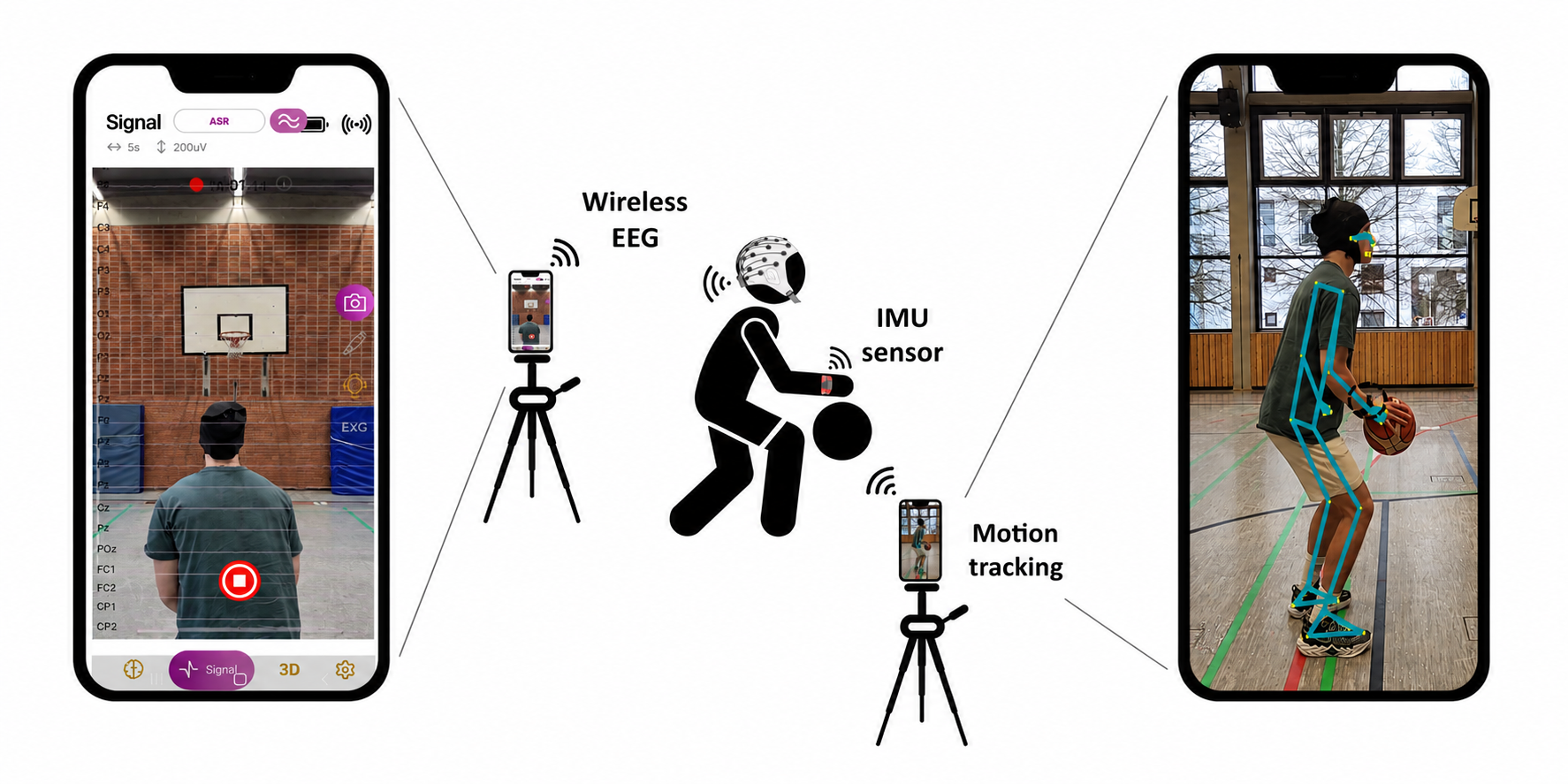
We assembled a portable setup based on two smartphones, a wireless 32-channel electroencephalography (EEG) system, and a small inertial measurement unit worn on the shooting wrist. One smartphone received the EEG signal and recorded video. The second captured body posture using camera-based pose landmark detection and recorded synchronized sensor streams via the Lab Streaming Layer framework embedded in custom apps.
Twenty-six experienced basketball players completed 120 free throws each across six blocks. They prepared and shot at their own pace, without an artificial cue instructing them when to begin. Preserving this natural timing was important for ecological validity, but it created a practical challenge: when does a free throw actually start?
Looking for the brain’s preparation signal
Our primary neural measure was the readiness potential, a slow negative shift in electrical brain activity that develops before a voluntary action. It has traditionally been studied with simple movements under controlled laboratory conditions. Detecting it during real basketball shooting would therefore provide a useful proof of principle for the portable recording setup.
At the group level, we observed the expected fronto-central negativity. At channel Cz, the readiness potential became progressively more negative during the final 400 milliseconds before movement onset. The scalp topographies, averaged across all participants, showed how this activity developed over fronto-central regions as movement onset approached.
This was the central result: a low-cost and lightweight setup using smartphones, wireless EEG, and a wrist sensor was sufficient to capture neural preparation together with whole-body movement outside the laboratory. The video abstract [Decoding the brain and body during basketball] is available for a visual overview of the study.
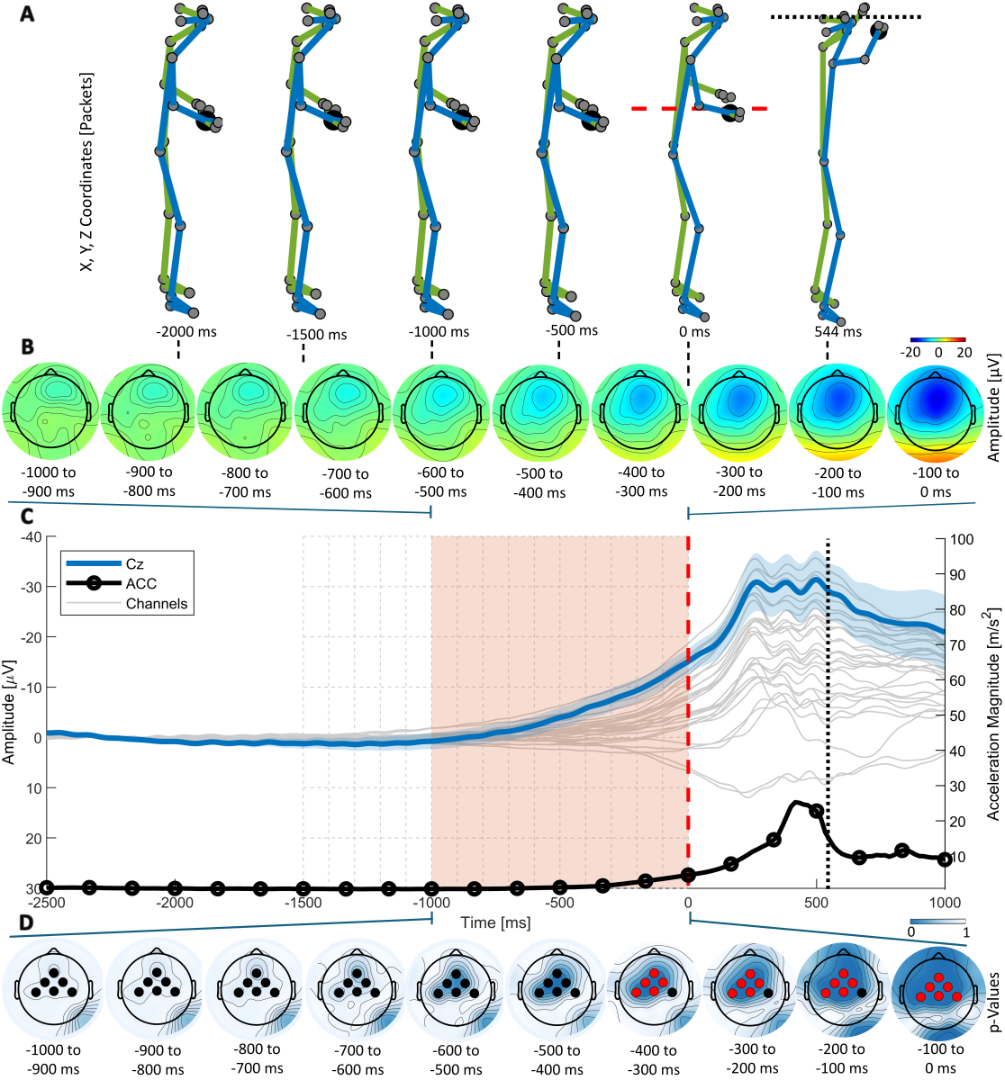
Grand average of simultaneous human motion capture and readiness potential. Body posture, wrist acceleration, and grand-average Event-Related Potential scalp topographies across participants illustrate the development of the readiness potential before movement onset. From Contreras-Altamirano et al. (2026), CC BY 4.0.
Brain and body signals unfolding together
The animation combines group-averaged body posture, grand-average ERP scalp topographies across participants, participant-level Cz activity, the grand-average Cz readiness-potential time course, and wrist acceleration relative to movement onset. Further demonstrations of the recording setup, motion tracking, and mobile applications are available in YouTube.
What the signal did—and did not—tell us
We also asked whether the readiness potential differed between successful and unsuccessful free throws. It did not. Across our analyses, readiness-potential amplitude was not reliably associated with whether a shot entered the basket. The maximum mean proportion of variance in shooting outcome explained by the neural features was only 4.7%.
This finding is scientifically important. Demonstrating that a neural signal can be recorded during natural movement is not the same as showing that the same signal predicts performance. A successful free throw depends on many interacting factors, and it would be misleading to expect one pre-movement brain measure to determine the outcome.
Finally, our exploratory pose analysis revealed participant-specific differences between successful and unsuccessful shots in 10 of the 26 players. These differences appeared at different body landmarks and time points, and the maximum mean proportion of variance explained by any pose landmark was 4.5%. The results therefore point more toward individual movement strategies than toward a universal posture for successful shooting.
Why the work matters beyond basketball
The main contribution of the study is methodological. Mobile brain/body imaging aims to study brain activity and behavior together in realistic settings. Our findings show that off-the-shelf smartphones and lightweight sensors can become part of a practical system for recording complex human actions.
Basketball was our test case, but the same general approach may support research on motor learning, rehabilitation, sports performance, and everyday behavior.
The open-source SENDA and RECORDA applications used in the acquisition framework are described in the iScience article Enhancing mobile brain and body imaging: Open-source solutions for real-world research applications, which presents further developments for synchronized mobile data streaming and recording.
The project website brings the study materials together, including the open-access paper, figures, the grand-average animation, demonstration videos, analysis code, and links to the acquisition applications. We hope that making the science openly visible will help other researchers evaluate, reproduce, and adapt the approach for their own real-world studies.
The broader lesson is that moving neuroscience out of the laboratory requires more than a portable EEG amplifier. Brain signals must be interpreted together with the body, the task, and the environment in which behavior occurs. By combining these elements, this study represents one step toward investigating brain-body dynamics where natural behavior actually happens and may help us better understand the neural mechanisms that support real-world action.
References and resources
Contreras-Altamirano, M., Klapprott, M., Jacobsen, N., Maanen, P., Welzel, J., & Debener, S. (2026). A portable solution for simultaneous human movement and mobile EEG acquisition: readiness potential for basketball free-throw shooting. Experimental Brain Research, 244, Article 153. https://doi.org/10.1007/s00221-026-07342-6
Debener, S., Minow, F., Emkes, R., Gandras, K., & de Vos, M. (2012). How about taking a low-cost, small, and wireless EEG for a walk? Psychophysiology, 49(11), 1617–1621. https://doi.org/10.1111/j.1469-8986.2012.01471.x
Gramann, K., Gwin, J. T., Ferris, D. P., Oie, K., Jung, T.-P., Lin, C.-T., Liao, L.-D., & Makeig, S. (2011). Cognition in action: imaging brain/body dynamics in mobile humans. Reviews in the Neurosciences, 22(6), 593–608. https://doi.org/10.1515/RNS.2011.047
Haupt, T., Maanen, P., Daeglau, M., Contreras Altamirano, M., Stritzke, A. S., Kiene, F., Welzel, J., Blum, S., Roheger, M., & Debener, S. (2026). Enhancing mobile brain and body imaging: Open-source solutions for real-world research applications. iScience, 29(7), 116647. https://doi.org/10.1016/j.isci.2026.116647
Jacobsen, N. S. J., Blum, S., Scanlon, J. E. M., Witt, K., & Debener, S. (2022). Mobile electroencephalography captures differences of walking over even and uneven terrain but not of single and dual-task gait. Frontiers in Sports and Active Living, 4, 945341. https://doi.org/10.3389/fspor.2022.945341
Klapprott, M., & Debener, S. (2024). Mobile EEG for the study of cognitive-motor interference during swimming? Frontiers in Human Neuroscience, 18, 1466853. https://doi.org/10.3389/fnhum.2024.1466853
Schurger, A., Hu, P. B., Pak, J., & Roskies, A. L. (2021). What is the readiness potential? Trends in Cognitive Sciences, 25(7), 558–570. https://doi.org/10.1016/j.tics.2021.04.001
Follow the Topic
-
Experimental Brain Research
This journal publishes original contributions on many aspects of experimental research of the central and peripheral nervous system.

Related Collections
With Collections, you can get published faster and increase your visibility.
Contributions of the reticulospinal system to sensorimotor control
The prominent role the reticulospinal system and descending reticulospinal pathway play in shaping spinal motoneuron excitability and the production of motor responses is being increasingly appreciated. One of the challenges of studying reticulospinal pathways in higher-order vertebrates (e.g. humans and non-human primates) is the difficulty in accessing these systems at the level of the brainstem.
This call of papers invites submissions with novel data showing the contributions of the reticulospinal system, or putative reticulospinal system, to the control of spinal motoneuron excitability, voluntary/involuntary movements, gait or postural control. These studies can include in investigations of the reticulospinal system in animal preparations or humans with or without neurological disorders. Submissions with innovative protocols for examining the contributions of reticulospinal systems to sensorimotor control are highly encouraged.
Publishing Model: Hybrid
Deadline: Ongoing
The contribution of muscle synergies to normal and abnormal motor control
The idea that the CNS may control complex interactions by modular decomposition has received considerable attention. Muscle synergies, computationally identified intermuscular coordination patterns, have been used to characterize neuromuscular control in humans. One important reason for the interest in understanding modularity in motor control is the impaired regularities in muscle activities in various pathologies. Investigations of muscle synergies, motor primitives, compositionality, basic action concepts, and related theoretical and experimental studies may contribute, at different levels, to advance our understanding of the modular architecture underlying motor behaviors. The goal of this Topical Collection is to present and discuss available evidence, conceptual frameworks, and basic issues related to intermuscular coordination patterns in motor control and learning. These cover a range of issues such as the effective dimensionality, neural underpinnings, movement invariants and biomechanical correlates, spinal maps of motoneuron activation, comparative physiology, development, and movement disorders. By bringing together researchers who are interested in understanding the building blocks of coordinating many muscles, we aim at promoting interactions between various research areas and techniques and providing a broad perspective on the notion of modularity in neuromuscular control.
Publishing Model: Hybrid
Deadline: Dec 31, 2026