Rescuing archived clinical CT for skeletal research
Published in Healthcare & Nursing and Social Sciences

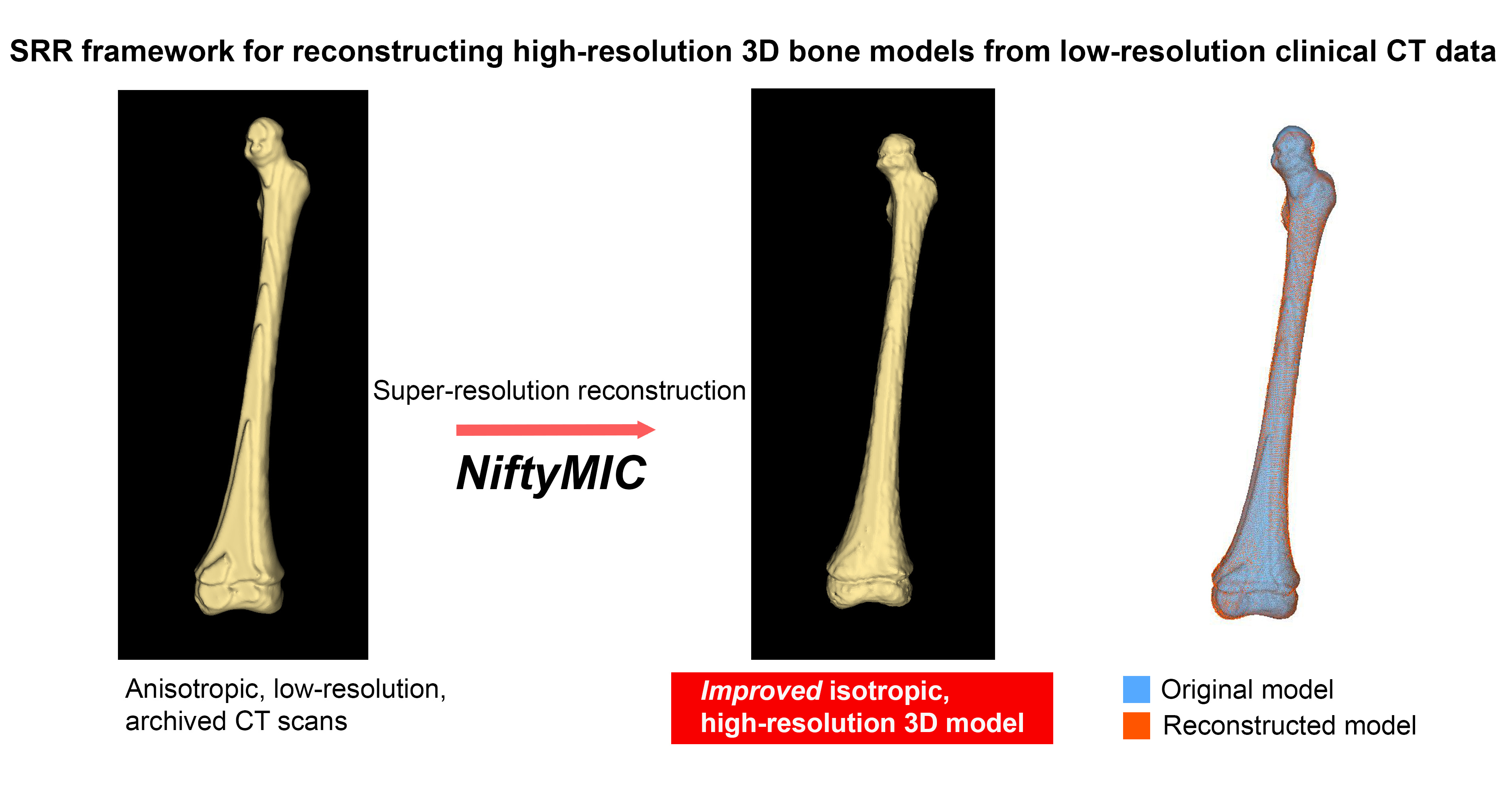
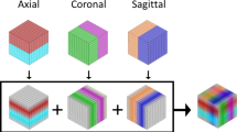

This project grew out of my dissertation data obtained from National Taiwan University Hospital. The plan was simple on paper: collect archived clinical CT scans of children’s long bones and build 3D models for osteometric research. In practice, the PACS rarely held the original isotropic volumes. What it held were multiplanar reconstructions: axial, sagittal, and coronal stacks with slices up to 5 mm thick. Models segmented from stacks like these look stepped and voxelated, and the metaphyseal margins in young children are often not resolved at any threshold. While smoothing can hide the stepping, but it also rounds off the very surface features that growth studies depend on. For subadult skeletal research, where dry bone collections are scarce, giving up on these archives was not a good option: We needed a way to improve the data without inventing it.
Super-resolution reconstruction is now dominated by deep learning, but those methods need large sets of paired high-resolution training data, which is exactly what retrospective clinical archives lack. Model-based SRR takes a different route. It treats each low-resolution stack as a geometrically transformed, noisy observation of one underlying high-resolution volume, and then estimates that volume by solving a regularized inverse problem. Three orthogonal stacks of the same bone, each sharp in a different direction, are a natural fit for this.
We used the open-source NiftyMIC framework, which was developed for fetal brain MRI. Most of its pipeline, including brain localization, motion correction, and slice-to-volume registration, was irrelevant for static CT of a single acquisition, so we used only the SRR component and supplied manually drawn masks to restrict the reconstruction to the target bone. We deliberately kept the developers’ default parameters and changed only the output voxel size. Our goal was to see whether a skeletal researcher could adopt the tool without becoming an image-processing specialist.
We reconstructed 61 long bones from 30 individuals aged 0 to 16 years and compared three versions of each bone: measurements from the hospital’s high-resolution PACS volume renderings, models from the original thick-slice stacks, and models from the SRR volumes. The SRR models agreed more closely with the PACS reference. Inter-observer error also fell, suggesting the smoother surfaces made landmarks easier to place consistently.
Geometrically, the two model types were broadly similar, with mean surface deviations within about 1 mm and a median Dice coefficient of 0.945. The larger local deviations clustered at the metaphyses, which is where the thick-slice models struggle most and where SRR made separating fusing epiphyses visibly easier. Our Bayesian mixed-effects models also showed that surface deviation increased with age, a pattern we think reflects how difficult the cartilage–bone interface is to resolve in developing skeletons on thick-slice CT.
The results suggest that thick-slice archives do not have to be a dead end. With freely available software and minimal parameter tuning, researchers can extract more usable skeletal data from clinical images that already exist, without additional radiation exposure or new scanning.
Follow the Topic
-
Scientific Reports
An open access journal publishing original research from across all areas of the natural sciences, psychology, medicine and engineering.

Related Collections
With Collections, you can get published faster and increase your visibility.
Cancer Prevention and Control
Publishing Model: Hybrid
Deadline: Feb 19, 2027
Healthy Aging
Publishing Model: Open Access
Deadline: Dec 31, 2026
