SLIMBRAIN Database: A Multimodal Image Database of In vivo Human Brains for Tumour Detection
Published in Healthcare & Nursing, Neuroscience, and Research Data

Surgery is still an unavoidable step in the treatment of most human brain tumours. Many of these tumours are not identifiable with the naked eye and there is little differentiation with normal tissue, which makes it difficult for neurosurgeons to identify them accurately. The challenge is to remove as much pathological tissue as possible while removing as little healthy tissue as possible, so as not to compromise the patient's quality of life. This is especially important when tumours infiltrate surrounding areas like glioblastoma multiforme. Researchers from the Universidad Politécnica de Madrid, in collaboration with neurosurgeons from the Hospital Universitario 12 de Octubre in Madrid, Spain, are trying to find a solution to this problem.

Traditional imaging techniques like magnetic resonance imaging and ultrasound are invaluable but have limitations, such as lower resolution or limited intraoperative applicability. In recent decades, however, a new approach has been used in medical research: hyperspectral imaging combined with artificial intelligence techniques.
Hyperspectral imaging captures a wide range of wavelengths beyond the visible spectrum, which can include infrared and ultraviolet. Unlike standard RGB images, which use three colour channels similar to what the naked human eye can see, hyperspectral images can include dozens or hundreds of spectral bands, each representing a specific wavelength. This richness of data allows for more detailed analysis of tissue composition, differentiating between different types of tissue based on their unique spectral signatures. Looking at the image below, on the left we have an RGB image synthetically created from all the wavelengths captured by one of the hyperspectral cameras used to create the SLIMBRAIN database. On top of this are 3 squares with pixels of different materials inside. By spectrally analysing the pixels inside, we can see on the right the so-called spectral signature, i.e. the behaviour of light across the spectrum for the different materials. As can be seen, each has a different behaviour, making it easier to distinguish between them, offering great potential for differentiating between healthy and pathological tissue.

Compared to traditional imaging approaches, hyperspectral imaging offers several advantages:
-
Non-Invasive: Unlike some imaging methods, hyperspectral imaging doesn't require contrast agents, making it safer for patients.
-
Non-Ionizing: No radiation is emited to the tissue, ensuring that no DNA is damaged and thus preserving tissue.
-
Real-Time Feedback: Surgeons can receive immediate information during surgery, aiding in precise decision-making, if the right equipment is used.
Hyperspectral images can obtain more information than we can see with our naked eyes, but they alone cannot delimit a tumour in the brain. In this context, an automated process is needed, which can be achieved by using sophisticated techniques that could account for biological differences between patients. This is why technologies based on artificial intelligence are being used to train models, which need a lot of data to differentiate pathological tissue from healthy tissue in the exposed brain. However, there is a lack of public hyperspectral data of in vivo human brains and the most common dataset available was captured with a hyperspectral camera uncapable of providing real-time feedback. That was one of the motivations why we are introducing the SLIMBRAIN database, to give researchers more data and to provide images from multiple cameras, enabling the development of real-time diagnostic tools.
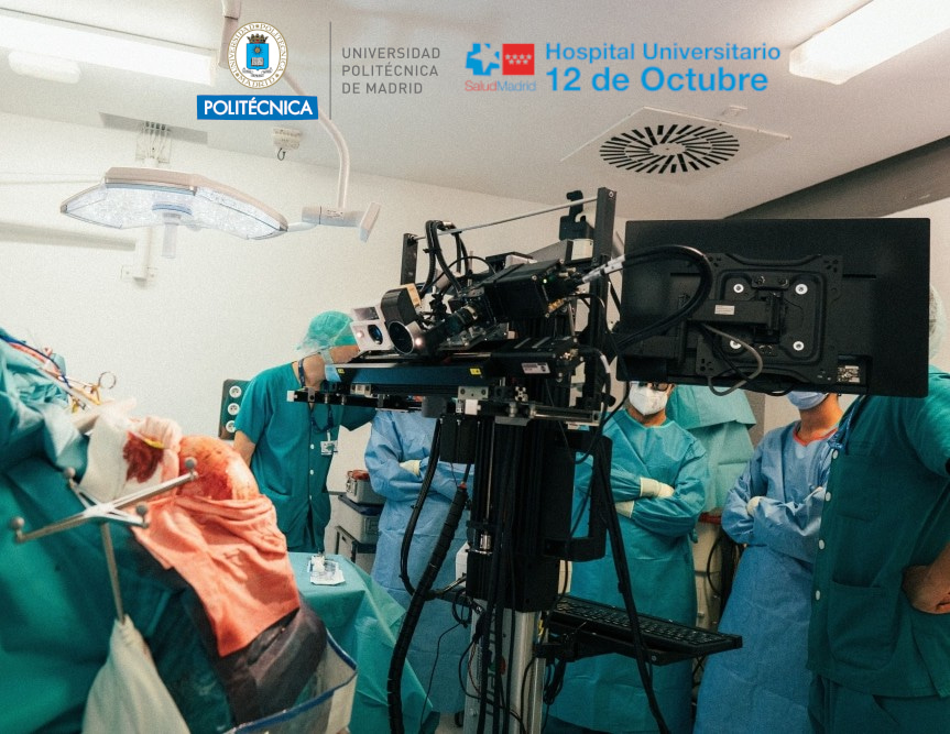
For the SLIMBRAIN database we have used two hyperspectral cameras: one with higher resolution but no video capability (linescan camera) and the other with lower resolution but with video capability (snapshot camera). The snapshot camera can provide live video of tumour delineation to the neurosurgeon, thus providing real-time solutions compared with the linescan camera, which is used to validate the performance of both cameras. These two cameras, among with other imaging and mechanical systems can be seen in the image below.

The SLIMBRAIN database holds data that can be used in various ways. For example, it can be used to train artificial intelligence models with over 1 million HS pixels, labelled by neurosurgeons, to reconstruct 3D scenes or to visualise RGB brain images with different pathologies. With artificial intelligence models, it is possible to classify different brain tissues from the hyperspectral images. These classifications often illustrate a coloured image to identify different tissues. However, thanks to the hyperspectral snapshot camera, it is possible to create real-time classifications of brain tissues. The video below shows on the left a hyperspectral video captured with the snapshot camera, whereas on the right the same video is classified by an artificial intelligence model with a specific colour mapping: green, blue, and pink for healthy, blood vessel, and meninges tissues, respectively. Note that in this example, the patient was suffering from an aneurysm so that no tumour is present on the surface.
Neurosurgeons usually rely on preoperative MRI scans - detailed images taken before surgery begins. These scans are extremely helpful, but there is a big problem: the brain does not stay exactly where it was after the skull is opened. This phenomenon is called brain shift, which refers to the natural movement or deformation of the brain that occurs during surgery after part of the skull has been removed. These changes mean that the structure of the brain can shift by several millimetres - or even more - which may not sound like much, but in brain surgery it is a great amount. It can mean that the MRI no longer matches the actual position of the brain during surgery. To overcome this, imaging systems are needed that work in real time, during surgery, and provide a 3D understanding of the brain's current state. But hyperspectral imaging alone is only superficial and 2D. That is why 3D information is critical, and that is another reason why the SLIMBRAIN database offers such 3D information through depth sensors, which measure how far or close areas are from the sensor. This is what makes the SLIMBRAIN database different from other public datasets. Moreover, by combining hyperspectral imaging with depth sensing technologies it is possible to generate 3D visualisations similar to the example below.
Magnetic resonance imaging is commonly used during neurosurgery to guide surgeons. It provides a detailed map of the brain before surgery begins. By matching real-time classification data from hyperspectral data with structural data from 3D depth images and magnetic resonance imaging scans, it would be possible to: update the magnetic resonance imaging map during surgery to account for brain movement; reconstruct a 3D model of the brain surface that reflects its current shape and condition; and provide neurosurgeons with a dynamic navigation tool that is far more accurate than a static image. Imagine that your magnetic resonance imaging scan is your detailed travel plan. But if a landslide blocks the road (brain shift), you need live GPS updates for a safe re-route. This is where hyperspectral imaging + 3D imaging + magnetic resonance imaging alignment could offer a huge potential tool for segmenting brain tumours in vivo.
🧠Access the SLIMBRAIN database🔎
🎓 Learn more about our recent research 📖
🔗 [2023] (Journal paper) SLIMBRAIN: Augmented reality real-time acquisition and processing system for hyperspectral classification mapping with depth information for in-vivo surgical procedures
🔗 [2024] (Journal paper) HyperMRI: hyperspectral and magnetic resonance fusion methodology for neurosurgery applications
🔗 [2024] (Preprint) LIBRA: Low spectral resolution brain tumor classifier for medical hyperspectral imaging
🔗 [2024] (Journal paper) Spectral analysis comparison of pushbroom and snapshot hyperspectral cameras for in vivo brain tissues and chromophore identification
🔗 [2025] (Journal paper) Unifying heterogeneous hyperspectral databases for in vivo human brain cancer classification: Towards robust algorithm development
Follow the Topic
-
Scientific Data
A peer-reviewed, open-access journal for descriptions of datasets, and research that advances the sharing and reuse of scientific data.

Your space to connect: The Psychedelics Hub
A new Communities’ space to connect, collaborate, and explore research on Psychotherapy, Clinical Psychology, and Neuroscience!
Continue reading announcementRelated Collections
With Collections, you can get published faster and increase your visibility.
Genomics in freshwater and marine science
Publishing Model: Open Access
Deadline: Jul 23, 2026
Genomes of endangered species
Publishing Model: Open Access
Deadline: Jul 01, 2026


Please sign in or register for FREE
If you are a registered user on Research Communities by Springer Nature, please sign in