The Virtual Radiologist for the 03:00 a.m. Transplant
Published in Sustainability, Computational Sciences, and Surgery

When considering how artificial intelligence is transforming medicine, the focus often defaults to how machine learning can enhance or surpass our current diagnostic methods. While this approach will undoubtedly drive improvements in patient care, arguably the greatest potential for machine learning in medicine lies in enabling innovative ideas that were previously unattainable—making the impossible, possible. It is this line of reasoning that was the motivating factor behind our study, “Improving prognostic accuracy in lung transplantation using unique features of isolated human lung radiographs”, recently published in npj Digital Medicine[1].
Transplant medicine is a highly specialized field where decisions about the suitability of donor organs must be made quickly, and often with limited information. This is particularly true for lung transplantation, where surgeons have only a few hours to assess organ viability. The combination of these tight time constraints and the complexity of the procedure has resulted in a situation where only a small percentage (~20%) of donor lungs are conservatively accepted for transplantation.
But this all changed in the early 2000s when a team of Surgeon-Scientists in Toronto, Canada developed the Toronto Ex Vivo Lung Perfusion (EVLP) system—an approach to sustain the lung outside of the body for a prolonged period of time. This groundbreaking biomedical technology has ushered in a new era for transplantation, enabling surgeons to postpone accept-reject decisions until they can observe how the lung functions under physiological conditions. EVLP can be seen as akin to a 'dry run' for the surgery, but with minimal risk to patients.
EVLP also offers unprecedented access to tissue samples and data from isolated human lungs, providing a unique opportunity in medicine to significantly enhance our understanding of lung function in a minimally invasive way. The success of the EVLP platform has also exponentially expanded the amount of data we can capture about a lung, meaning we must now work to extract valuable information from this vast new resource at our fingertips.
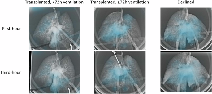
In 2023, our team was the first to explore EVLP data using complex mathematical models and machine learning. We showed that a model, termed InsighTx, that was trained to interpret the physiological, biological and biochemical data generated during EVLP was highly accurate in identifying suitable donor lungs for transplantation [2]. This work set the stage for standardized decision-making during EVLP by leveraging the largest known dataset of isolated lung function captured by the Toronto Lung Transplant Program over the past 15 years. However, the foundational InsighTx model did not include imaging. Though we routinely collected two radiographs of every lung placed on the EVLP system after one- and three-hours of perfusion, in our first iteration of InsighTx we needed to carefully consider how a machine learning tool would be used in practise and ensure that it would fit seamlessly into the clinical workflow. Images presented unique challenges—they weren't routinely collected by all EVLP centres and, more importantly, radiologists weren't typically involved as part of the standard EVLP team. So what do you do when you have a powerful source of information that is not readily accessible to clinical teams? You develop a new model that can incorporate EVLP X-ray images in an accessible way.
Our study builds on two previous reports: (1) the development and validation of the InsighTx model [2]; and (2) demonstration that EVLP radiographs are clinically meaningful [3]. The latter study used a ‘traditional’ methodology where a fellowship-trained radiologist manually reviewed and scored hundreds of images for the study. Conversely, the central goal of our latest work was to use a neural network-based approach to read and extract important features from the lung X-rays. By using machine-learning we could envision a more practical scenario where the entire process of image analysis is automated, thereby obviating the need for a radiologist to be present for every EVLP—including the ones at 03:00 a.m. It's important to note that our approach wasn’t about replacing existing personnel on the clinical team, but instead adding expertise to the team at no additional cost, through unlocking data for clinical decision-making that wasn't previously available.
In order to achieve this goal, we followed a pragmatic approach that started with pretraining our model on large, publicly available datasets and then fine-tuning our model on over 1,000 EVLP X-ray images. The most important aspect of our study was to investigate whether convolutional neural network (CNN)-based image features would significantly improve the performance of the foundational InsighTx model. In showing that this indeed was the case, we knew that the information ascertained by the neural network was a key building block for future updates of InsighTx. Perhaps most importantly, the image pipeline can be fully automated to truly provide this data to clinical teams at any time to enhance their assessment of a donor lung on the EVLP system.
As promising as our results were, we were keenly aware of the inherent risks of blindly trusting a machine leaning algorithm. To that end, it was important for us to investigate how and what the model was ‘looking at’ when extracting image features. For this, we were able to draw expertise from our radiology team and their manually derived labels. We were able to show that the components of the images identified using machine learning were aligned with findings in anatomical locations that were clinically sensible. Moreover, there were some CNN-derived image features that did not correlate with manual labels. Taken together, this mix of known and unknown image components was extremely reassuring to our team as it meant that the model was able to not only identify image findings (i.e., consolidation and infiltration) that we know to be clinically relevant, but also patterns that were not obvious to the human eye— thus showing the incredible strength of the machine learning approach to medicine.
Machine learning is a powerful tool for advancing our understanding of health and disease. Focusing on applications that are only possible using artificial intelligence opens the door to exciting and innovative projects, such as the recent work with radiographic images of isolated human lungs. The resulting tools and techniques hold the greatest potential for improving patient care.
Follow the Topic
-
npj Digital Medicine

An online open-access journal dedicated to publishing research in all aspects of digital medicine, including the clinical application and implementation of digital and mobile technologies, virtual healthcare, and novel applications of artificial intelligence and informatics.
Related Collections
With Collections, you can get published faster and increase your visibility.
Evaluating the Real-World Clinical Performance of AI
Publishing Model: Open Access
Deadline: Jun 03, 2026
Impact of Agentic AI on Care Delivery
Publishing Model: Open Access
Deadline: Jul 12, 2026


Please sign in or register for FREE
If you are a registered user on Research Communities by Springer Nature, please sign in