Building the human alveolar epithelium in vitro: why achieving developmental stages matters
Published in Bioengineering & Biotechnology and Cell & Molecular Biology

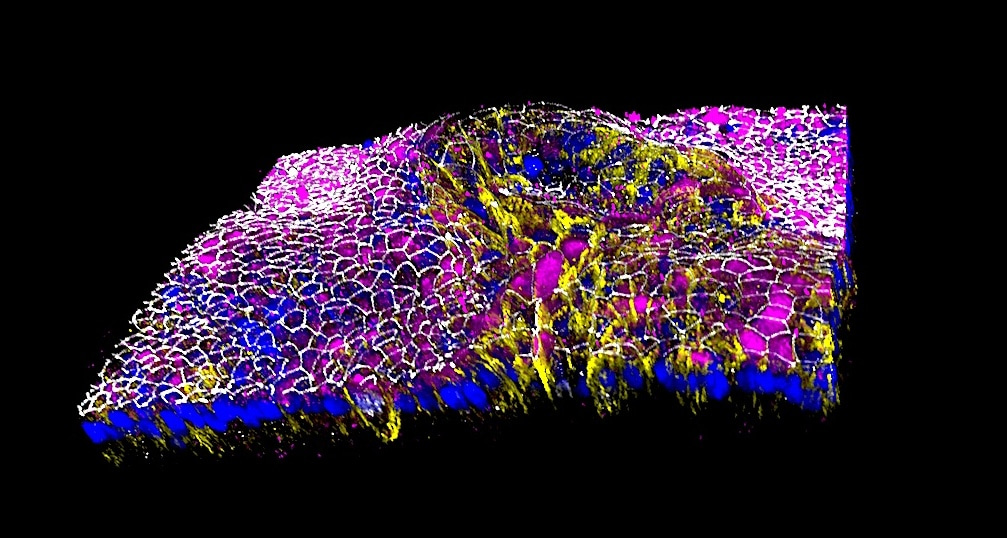

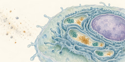
The alveoli are tiny air sacs where oxygen enters the bloodstream and carbon dioxide is removed. Their surface consists mainly of thin alveolar type 1 (AT1) cells, which enable gas exchange, and alveolar type 2 (AT2) cells, which produce surfactant, a mixture that prevents the alveoli from collapsing. During fetal development, both cell types arise from shared lung progenitors.
Patient-specific induced pluripotent stem cells can be used to model this process for studying lung development, disease, drug responses, and potential regenerative therapies. However, generating cells that express familiar markers is only part of the challenge. Because the same marker can appear at multiple developmental stages. Cells described as “fetal,” “immature,” or “mature” may therefore represent distinct developmental states rather than simply incomplete versions of adult cells. Therefore, recognizing these states is important for modeling diseases that arise at different periods of life.
Many groups have generated iPSC-derived AT2 organoids, or “iAT2” organoids, to study disorders associated with the surfactant genes SFTPC and SFTPB, as well as pulmonary fibrosis. AT2-like cells can also be guided toward an AT1-like identity through pathways such as Hippo-YAP1, which helps cells respond to stretching and changes in shape. Yet lung development involves more than biochemical signals.
Developing lung cells interact with surrounding fibroblasts, experience repeated stretching, and eventually encounter air. We therefore asked which developmental states current “iAT2” organoids represent and whether fetal lung fibroblasts, mechanical stretching, and air exposure could guide them toward more specialized alveolar identities.
Finding an unexpected developmental identity
We first improved the production of fetal lung progenitors (FLPs) from iPSC-derived definitive endoderm. These progenitors provide a consistent starting point for generating the epithelial cells that eventually form the alveoli.
We initially expected that SFTPC-expressing organoid cells could be classified as “AT2 cells”. However, as we examined the cells more closely, the story became less straightforward. They also expressed the AT1-associated marker AGER and contained fewer, less-developed lamellar bodies, which store and release surfactant. A turning point came when we compared our organoids with primary human fetal lung tissue. Their broader gene and protein profiles suggested that, rather than fitting neatly into an AT2 identity, the cells more closely resembled late tip and late stalk populations found in the fetal lung at approximately 15 to 18 weeks of gestation. These developing populations retain the potential to form either AT1 or AT2 cells. This finding changed how we interpreted our organoids.
Stem-cell differentiation is often evaluated by the appearance of hallmark genes associated with key developmental milestones. Although these markers are useful, cells do not all differentiate at the same rate or follow the same path. They may also pass through temporary developmental states before committing to a specialized identity. SFTPC is an important AT2 marker, but it is also expressed in earlier developmental states including late tip and late stalk. This distinction has practical consequences. Measuring differentiation by SFTPC alone could therefore mistake a progenitor population for more specified AT2 cells. By examining broader gene and protein profiles, cellular structure, and developmental potential, we could ask not only whether the cells expressed SFTPC, but also which developmental state they had reached and what they could still become.
Adding signals for later alveolar development
Once we recognized the developmental identity of the organoids, we asked whether we could guide them toward later fetal alveolar states. We added human fetal lung fibroblasts, supporting cells that help shape the epithelial environment through developmental signaling pathways, including fibroblast growth factors, Notch, and Sonic Hedgehog. Based on previous studies, we also inhibited LATS1 to increase YAP1 activity and support fetal-like AT1 differentiation. Active YAP1 enters the nucleus, where it regulates genes associated with mechanical responses and AT1 development. Together, these interventions promoted fetal-like AT1 and AT2 populations, resembling those found at approximately 18 to 20 weeks of gestation.
This was a step forward, but some cells continued to express both AT1- and AT2-associated markers, suggesting that their identities remained incompletely resolved. Fibroblast co-culture and biochemical activation of YAP1 alone were therefore insufficient to consistently guide cells toward distinct alveolar states. A challenge for future studies is to identify the additional signals that help developing cells complete and maintain their commitment to either an AT1 or AT2 identity. This led us to consider an important feature missing from conventional organoid cultures: the physical environment of the developing lung.
Bringing cyclic movement and air into the model
Before birth, fetal breathing movements repeatedly stretch the developing tissue. Around birth, lung fluid is cleared, and the alveoli is exposed to air. After birth, continuous cycles of breathing create mechanical forces that influence cell shape, signaling, and function.
We therefore tested whether cyclic stretch and air exposure could advance the cells beyond their fetal-like state. We first stretched the cells while submerged, then used an air-liquid interface that exposed one side to air while the other remained in a nutrient-containing medium.
Air-exposed cyclic stretch activated Hippo-YAP1 signaling and reduced the number of cells with co-expressing AT1 and AT2 markers. The cells also became larger and thinner, consistent with AT1 development, while pro-SFTPC levels increased above primary fetal lung levels but remained below adult lung levels. Together, these changes suggested more specialized alveolar features.
These findings suggest that supporting cells, mechanical stretch, and air exposure may provide complementary signals that help shape alveolar cell identity and function.
Building a developmental roadmap for future PSC models
Our study systematically tested how biochemical, cellular, and physical conditions influence alveolar development in vitro. These included changing the culture medium, manipulating mechanosensitive pathways with small molecules, adding fetal lung fibroblasts, and exposing the cells to mechanical stretch and air. Although these conditions promoted more specialized and mature-like AT1- and AT2 features, they did not recreate a fully mature human alveolus. This limitation reflects the complexity of developing the lung. Important pieces of the puzzle remain missing from our model, including interactions with immune and endothelial cells, vascular structures, and the shear forces generated by blood flow. Incorporating these components may help us better understand how different signals work together to shape alveolar development.
This work emphasizes a more practical question: What developmental stage are we modeling with current platforms/methods, and is it appropriate for the intended use? Answering this could improve the selection and interpretation of cells used to model diseases arising at different periods of life. It could also guide whether stem-cell-derived cells are ready for transplantation or require further expansion and maturation.
Our work therefore emphasizes a practical question: What developmental stage are we modeling, and is it appropriate for the intended application? Answering this question could improve how cells are selected and interpreted for modeling diseases that arise at different stages of life. It could also help determine whether PSC-derived cells are suitable for transplantation or require further expansion, differentiation, or maturation.
Rather than defining cell identity through a few familiar markers, our findings support examining molecular profiles, cellular structure, and function within a developmental context. Comparisons with third trimester and postnatal lung tissues will be especially important for determining how closely these cells resemble those in the perinatal lung, although suitable reference datasets remain limited.
Next, the challenge is to identify which stages of lung development our models capture, where they remain incomplete, and which additional signals may guide cells toward more specialized and functional states.
Follow the Topic
-
Communications Biology
An open access journal from Nature Portfolio publishing high-quality research, reviews and commentary in all areas of the biological sciences, representing significant advances and bringing new biological insight to a specialized area of research.

Related Collections
With Collections, you can get published faster and increase your visibility.
Healthy Aging
Publishing Model: Open Access
Deadline: Dec 31, 2026
Cell death and inflammatory signalling
Publishing Model: Hybrid
Deadline: Jan 28, 2027