Chlamydia trachomatis: bad reputation with a fascinating story.
Published in Microbiology, Protocols & Methods, and Cell & Molecular Biology

Most people know Chlamydia trachomatis as cause of sexually transmitted infections. But behind its bad reputation as troublesome disease lies an organism with an extremely interesting biology.
The Ultimate Nutrient Thief
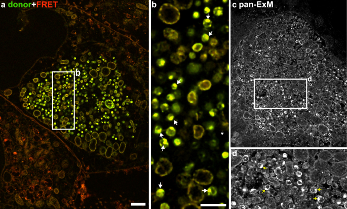
The bacterium enters the cells of our body and generates its own replicative compartment, the so-called inclusion. These inclusions are fascinating structures densely packed with bacteria and so huge that they occupy a significant amount of space within the cell, often becoming larger than the host nucleus (see Figure 1, a and c). Interestingly, C. trachomatis lost its ability to replicate without a host cell and is therefore highly dependent on host metabolites that are hijacked from human cells during the infection. One important type of metabolite are sphingolipids, a class of highly abundant membrane lipids that are taken up from the host and incorporated into bacterial membranes (see Figure 1, c and d).
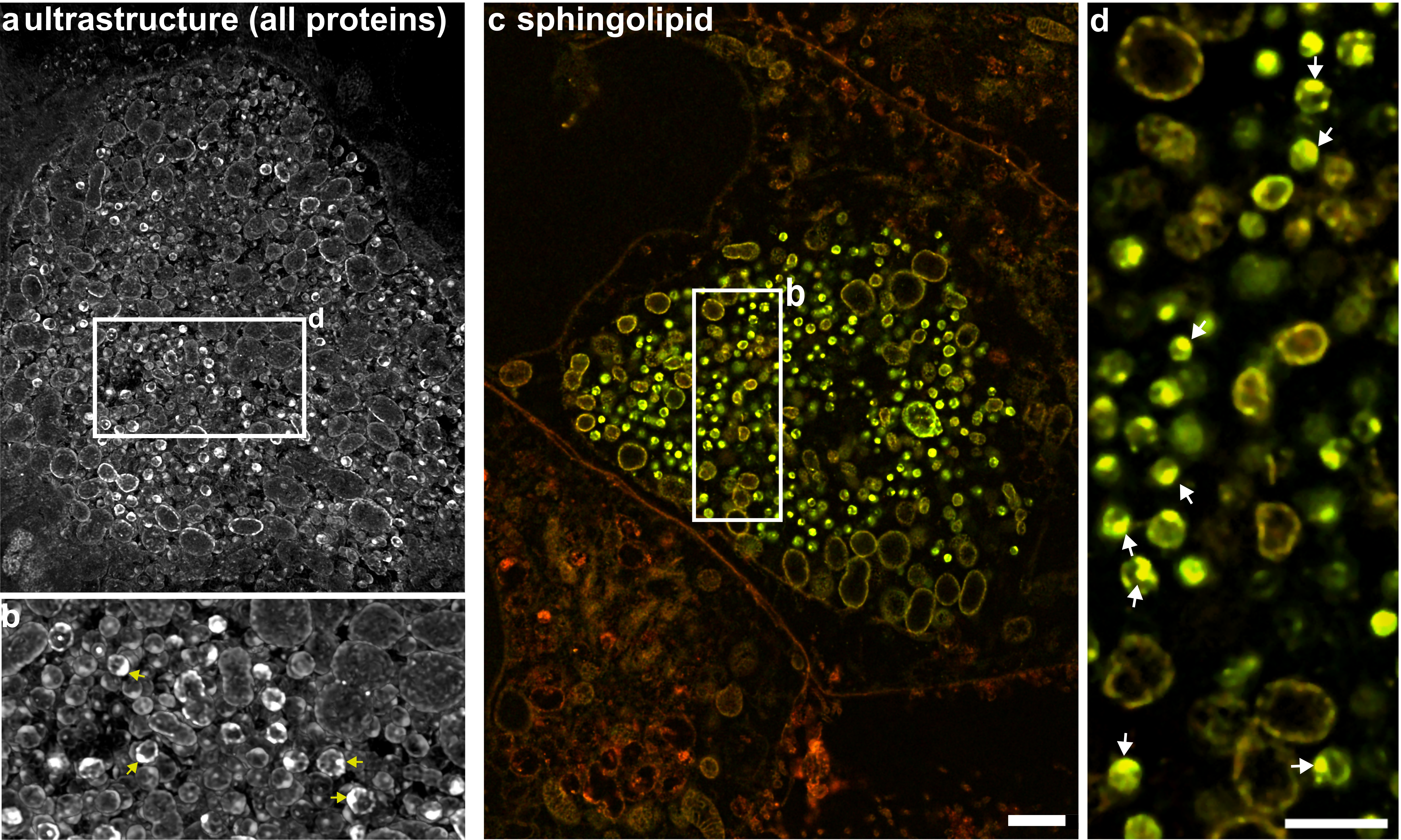
Figure 1 C. trachomatis inclusions visualized by super-resolution expansion microscopy (ExM). C. trachomatis inclusions were stained with an N-hydroxysuccinimid ester (a, b) that labels all proteins and visualize the ultrastructure or a sphingolipid analog (c, d). Scale bar: 1 µm.
A Shapeshifting Lifecycle
Moreover, C. trachomatis switches between two distinct developmental forms: elementary bodies (EBs): The tough, infectious form that survives outside human cells. In this inactive stage, the bacterium tightly packs its DNA into a dense core called the nucleoid to protect its genetic code and reticulate bodies (RBs): The active, reproducing form inside host cells.
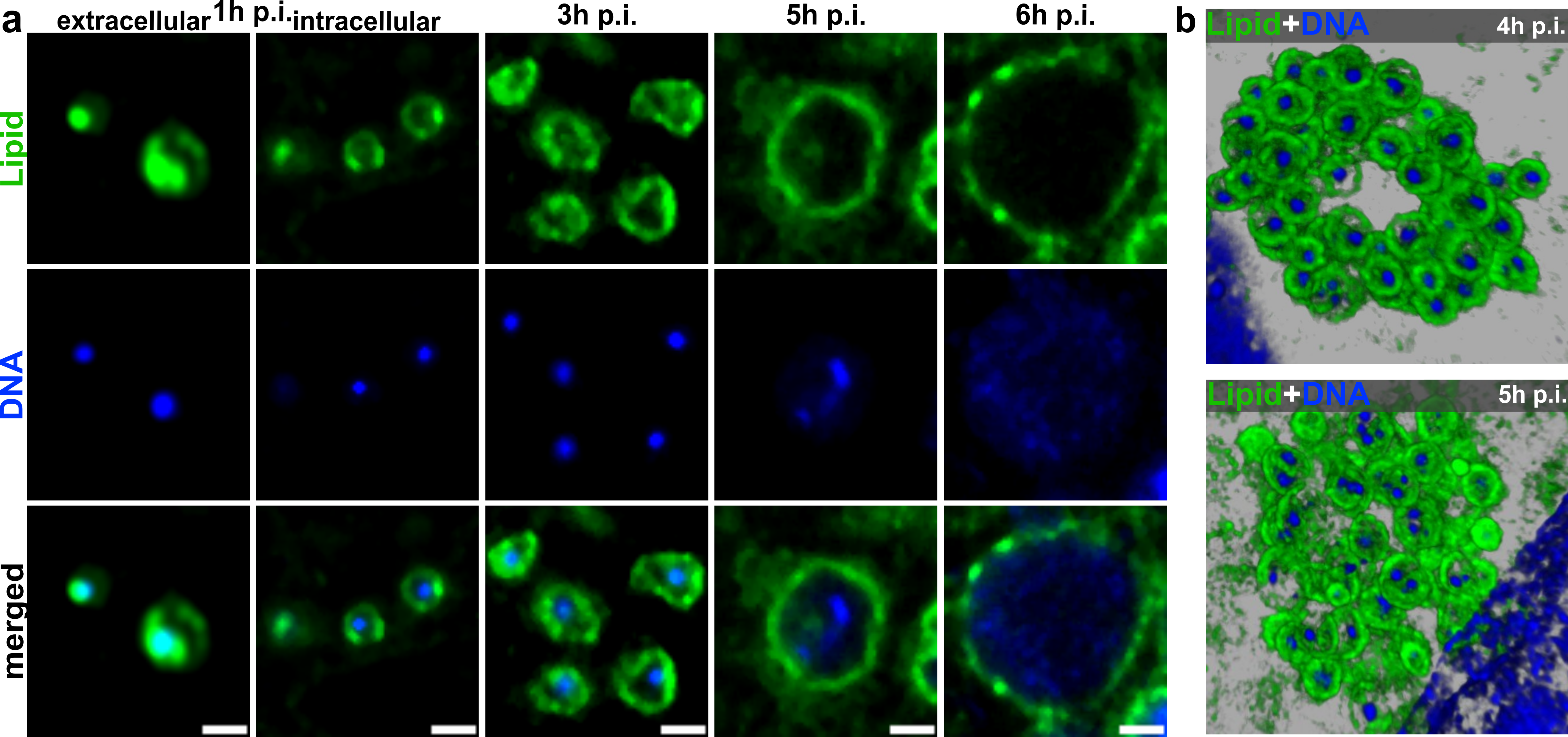
As soon as the EB has entered the host cell, a differentiation process is initiated, where the nucleoid is unwrapped and the EBs transform into RBs that then replicate and establish the inclusion (Figure 2, a and b).
Too Small to Catch?
Although these characteristics make C. trachomatis infections particularly interesting to study, they also impede any investigation with spatial resolution. Conventional light microscopy techniques possess a diffraction limit of 250 nm. The diminutive size of EBs, only measuring 200-300 nm, places them under this resolution limit. This led to the long-held belief that C. trachomatis is a virus, before it was recognized as bacterium in the 1960s. Moreover, the resolution of conventional light microscopy is often not sufficient to distinguish individual bacterial particles in the highly crowed inclusion.
Expansion Microscopy (ExM), developed by Edward Boyden in 2015, circumvents this resolution limit with a clever trick: instead of zooming in with a better microscope, you simply make the sample itself bigger. Therefore, a specimen is embedded into a swell-able hydrogel that upon hydration expands to 4×, 8× or even 20× of its original size. Ultimately, this results in an enlarged sample which enables a higher spatial resolution achievable on conventional light microscopes.
You might wonder: why bother with ExM when electron microscopy delivers even sharper images? In ExM, biomolecules can easily be tagged with fluorescence dyes. It also facilitates 3D reconstructions by simply recording z stacks with a confocal microscope (see Figure 1 a, b and Figure 2, b) and importantly, it brings high-resolution imaging to standard light microscopes, which are readily available in most research labs.
Applied to C. trachomatis, ExM drastically sharpens our view on inclusions and the tiny EBs. 8×ExM even facilitates the identification of structures, such as the nucleoid, within individual particles. On conventional microscopes, these tiny features normally get squished into a single pixel!
Uncovering the hidden DNA-Lipid Interface
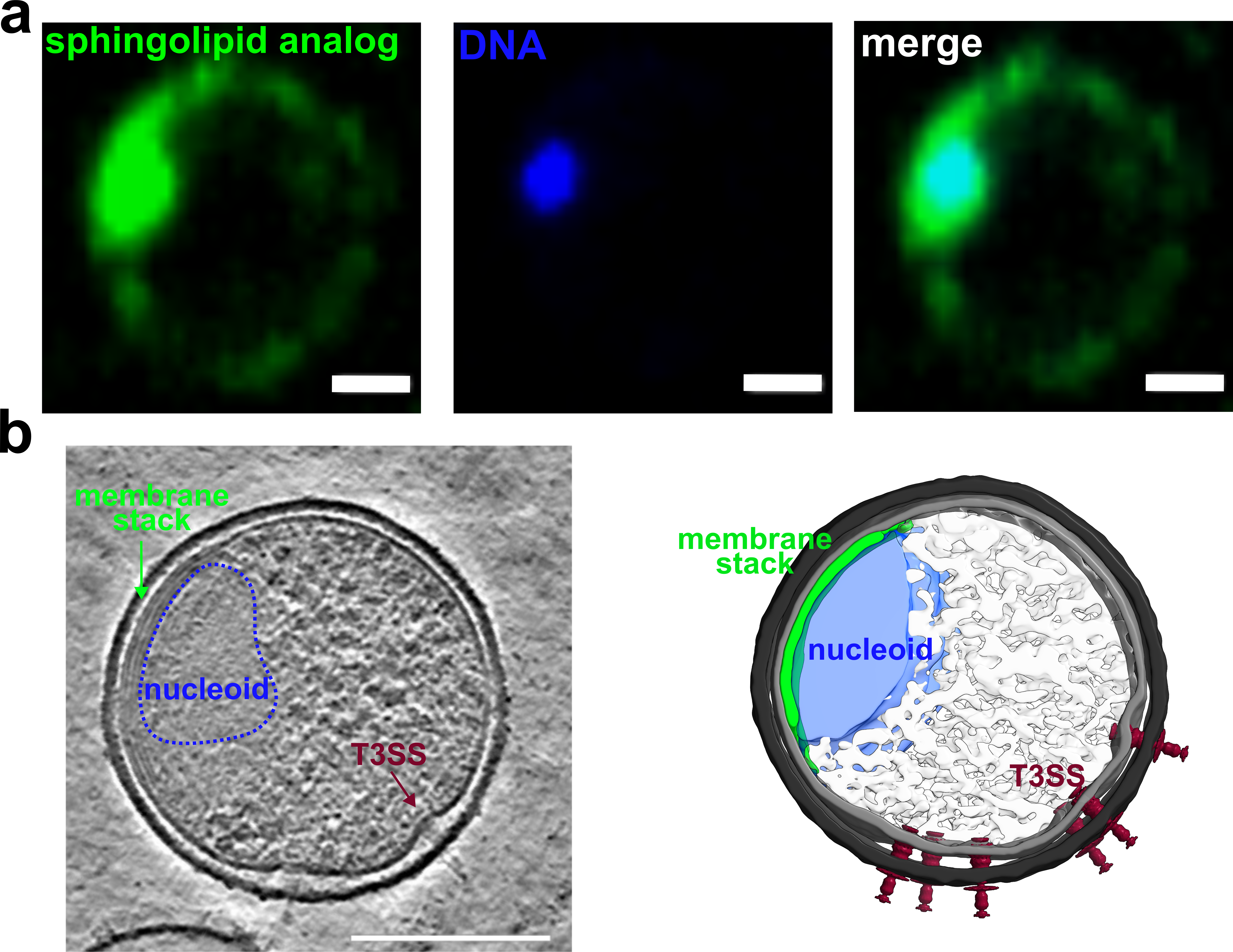
By using 8×ExM to visualize DNA and lipids, we discovered something unexpected: a synthetic sphingolipid analog is incorporated into the nucleoid of EBs (Figure 3, a). In our images, this can be seen by the round protrusion in the lipid channel that perfectly overlaps with the DNA signal, which marks the nucleoid of the particle. As reference, we also recorded EBs by cryo electron tomography, where the particles are flash frozen and afterwards analyzed with an electron microscope. This revealed a membrane stack to which the DNA-containing nucleoid is tethered (Figure 3, b). While cryo electron tomography is ideal to study the native structure of EBs, it does not allow specific staining for DNA and lipids. Because of this, combining both methods is essential: ExM shows us what molecules are there, while cryo electron tomography reveals more details about the native ultrastructure.
Watching the Transformation Unfold
When the infectious EBs start transforming into replicating RBs, something remarkable happens first: the lipids are released from the nucleoid, and the nucleoid detaches from its membrane stack (Figure 2, a). We can watch this step-by-step transition unfold. By comparing extracellular EBs (1 h p.i. “extracellular”), that have not initiated the differentiation process, with intracellular ones that have just begun their transformation (1 h p.i. “intracellular”), we can clearly see DNA and lipids separating as the infection proceeds.
Later in the infection, when RBs again redifferentiate into EBs, sphingolipids are massively metabolized in the inclusions of C. trachomatis. This we could investigate by our sphingolipid analogs and a trick that made it possible to trace their metabolization with spatial resolution and ExM. The metabolized lipids are then packed into the membrane stack and the nucleoid of EBs.
The Limits of Resolution—And What's Next
Even with 8×ExM we reached a resolution limit of ~30 nm. While that is impressively sharp, it is not good enough to reveal the exact architecture where lipid meets DNA. Because the DNA inside the nucleoid is so tightly packed, and because electron microscopy lacks specific lipid dyes, capturing this exact structure remains a challenge. For now, the fine details of this lipid-DNA interface remain a fascinating mystery that needs to be determined in future studies.

Follow the Topic
-
Nature Communications
An open access, multidisciplinary journal dedicated to publishing high-quality research in all areas of the biological, health, physical, chemical and Earth sciences.

Related Collections
With Collections, you can get published faster and increase your visibility.
Women's Health
Publishing Model: Hybrid
Deadline: Ongoing
DNA repair and human disease
Publishing Model: Hybrid
Deadline: Oct 31, 2026