Cracking the code: Using deep learning to study bone fracture micro-mechanics and toughness
Published in Electrical & Electronic Engineering

What do biologists, mechanical engineers, and computer scientists have in common? More than you might think. In 2019, our lab wanted to solve a perplexing "catch-22" in the realm of bone imaging: how do we perform near real-time mechanical testing and computed tomography (CT) without exposing our bone samples to excess radiation damage?
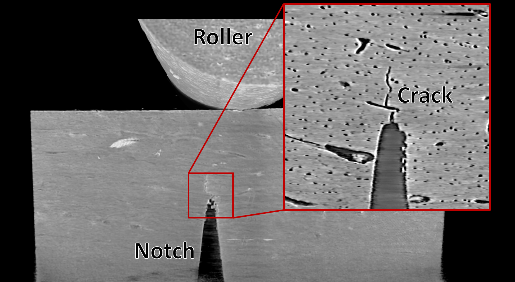
In this project, we had two aims: 1) to measure how well bone resists fracture using a toughness test and 2) to capture images of bone's internal structure and the shape of the crack during fracture using micro-CT. These two aims have competing interests. If the bone in the toughness test is imaged repeatedly using micro-CT, the toughness results will become unreliable due to radiation exposure's harmful effects on bone mechanics. Damage due to radiation is especially a problem when using synchrotron radiation micro-CT (SRμCT) imaging, which uses high flux x-rays to image at a microscopic scale.
Theoretically, an in situ test such as the one described above could provide extraordinary insight into the nature of bone fracture and how cracks propagate through bone at the microscale. If we could track the deformations in bone during multiple steps of crack growth, we could relate collagen damage due to disease to the crack path, deformation behavior, and toughness. Because the mechanics of bone fracture as it relates to disease could help inform effective therapies and treatments in the future, we wished to solve this "catch-22" of radiation damage in our tests.
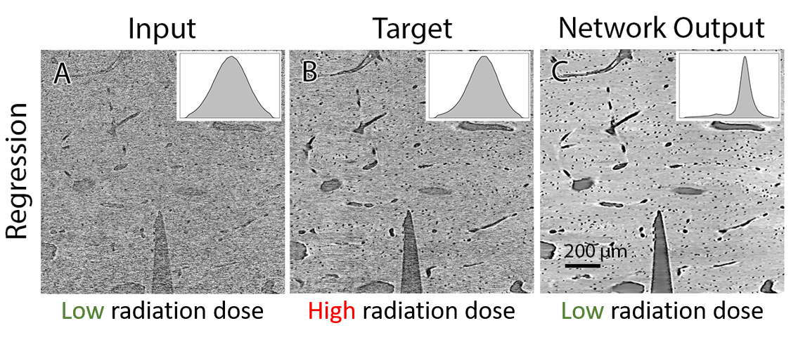
We leveraged deep learning to circumvent the radiation damage and perform the study. We reduced the damage caused by radiation exposure by taking lower-quality images of the bone during the toughness test. Subsequently, we trained a neural network to denoise these low-quality images based on high-quality target images, allowing us to study toughness and crack propagation in bone without damaging it (Figure 1). The development of this technique required us to tackle these challenges from the perspectives of biology, mechanical engineering, and computer science.
What did we learn from the in situ technique from a biological perspective?
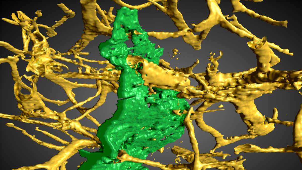
The biological aspect of studying collagen damage in bone is both the beginning and final goal of this project. To mimic the effect of collagen damage in disease, we induced collagen damage by heat-treating bone at varying temperatures to provide a gradient of collagen damage. During testing, we found that increased collagen damage at the nanoscale influenced the strain state and crack path at the microscale and decreased toughness at the macroscale. We hope to apply this technique to bone disease to discover how these diseases impact bone fracture mechanics to inform potential treatments.
What did we learn from the in situ technique from a mechanical engineering perspective?
Using the in situ testing technique, we found the impact of collagen damage on toughening mechanisms in bone. With more damage to collagen, we found that a growing crack changed directions fewer times. Changing directions, or deflecting, during crack growth is an important, natural way for a propagating crack to dissipate energy. With more damage, we found this natural behavior was hampered, accompanied by a drop in fracture toughness. Understanding the project from a fracture mechanics and solid mechanics background was integral to connecting our findings back to the biological bottom line.
How can we develop the in situ testing technique from a computer science perspective?
Although the denoising performed by the neural network in this work is not part of the final results, it is the glue that holds the project together. Without it, the original "catch 22" of in situ micro-CT testing and radiation damage would remain unsolved. We collaborated with Daniël M. Pelt from the Netherlands to use his mixed-scale dense network architecture for this project to denoise our low radiation dose images (Figure 2). In our initial work, we found that this architecture out performed other denoising neural networks. It was necessary to have a strong background in computer science to understand how the network functioned and how best to apply it to our data.
Let's go back to our initial question, "what do a biologist, mechanical engineer, and computer scientist have in common?" They are the same person in this work, and we have a lab full of them. To solve intricate problems, combining several fields of research is often required. Here, we found through a new in situ toughness testing technique that collagen damage at the nanoscale reduces crack deflection at the microscale and lowers toughness at the macroscale.
Follow the Topic
-
Communications Materials
A selective open access journal from Nature Portfolio publishing high-quality research, reviews and commentary in all areas of materials science.

Related Collections
With Collections, you can get published faster and increase your visibility.
Materials for quantum sensing and computing
Publishing Model: Open Access
Deadline: Jul 09, 2026
Lead-free perovskite solar cells
Publishing Model: Open Access
Deadline: Jul 08, 2026


Please sign in or register for FREE
If you are a registered user on Research Communities by Springer Nature, please sign in