Disc Degeneration and Facet Joint Osteoarthritis: What Does kMRI Reveal at L5/S1?
Published in Neuroscience, Zoology & Veterinary Science, and Surgery
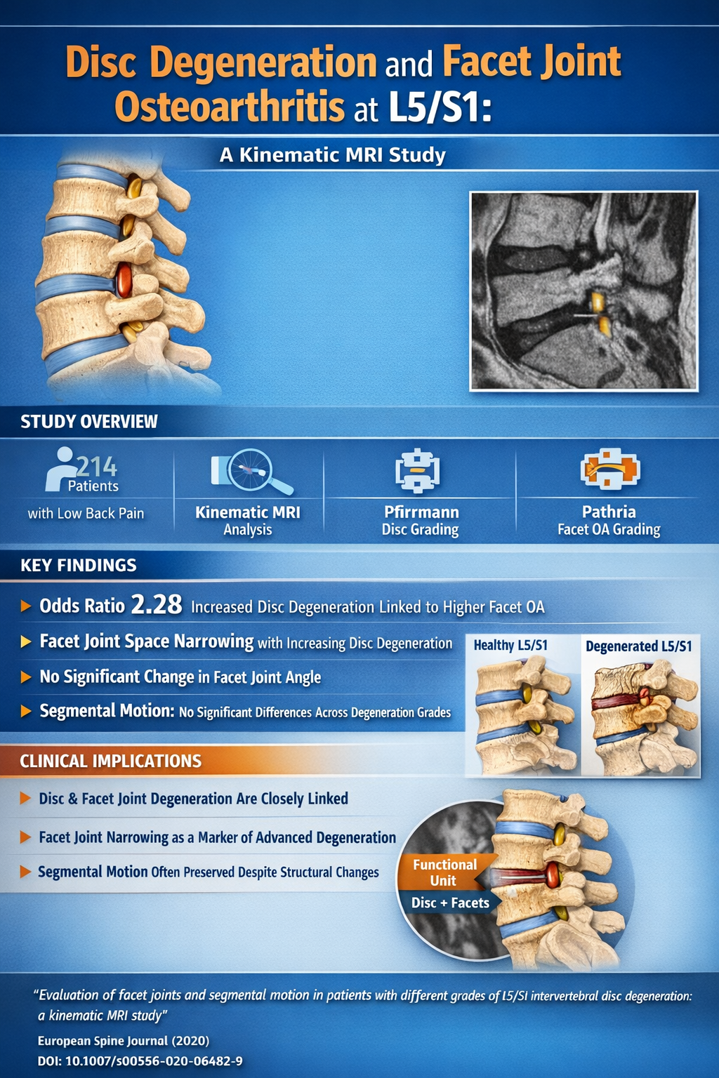
Low back pain is often the result of complex, interrelated degenerative processes. While intervertebral disc degeneration and facet joint osteoarthritis are frequently discussed together, their dynamic relationship and biomechanical implications remain incompletely understood.
In our study published in European Spine Journal, we used kinematic MRI (kMRI) to explore how different grades of L5/S1 disc degeneration relate to facet joint changes and segmental motion.
🔎 Study Overview
- 214 patients with low back pain
- Retrospective analysis using kMRI
- Grading systems:
- Disc degeneration: Pfirrmann classification
- Facet joint osteoarthritis: Pathria classification
- Evaluated:
- Facet joint angle and joint space width
- Segmental angular and translational motion
📊 Key Findings
• Strong association between disc degeneration and facet OA
– Odds ratio: 2.28 (P = 0.008)
• Positive correlation between degeneration severity
– Higher Pfirrmann grades correlated with higher facet OA grades
• Facet joint space narrowing with degeneration
– Significant decrease in joint space width with increasing disc degeneration
• No significant change in facet joint angle
• Segmental motion (angular & translational)
– No significant differences across degeneration grades
💡 Clinical Takeaway
This study reinforces the concept that disc degeneration and facet joint osteoarthritis are closely linked degenerative processes, particularly at the L5/S1 level.
However, despite structural degeneration:
➡️ Segmental motion may remain relatively preserved, challenging the assumption that increasing degeneration always leads to measurable instability.
⚖️ What This Means for Practice
- Degenerative changes should be viewed as a functional unit (disc + facets) rather than isolated findings
- Facet joint space narrowing may serve as an additional imaging marker of advanced degeneration
- kMRI provides valuable dynamic insights beyond static imaging
📄 Evaluation of facet joints and segmental motion in patients with different grades of L5/S1 intervertebral disc degeneration: a kinematic MRI study
European Spine Journal (2020)
DOI: 10.1007/s00586-020-06482-9
Mohamed Kamal Mesregah , Haiyin Lee, Sidney Roberts, Carson Gardner, Ishan Shah, Ian A Buchanan, Changqing Li, Zorica Buser, Jeffrey C Wang
Article links:
https://pubmed.ncbi.nlm.nih.gov/32504265/
https://link.springer.com/article/10.1007/s00586-020-06482-9
I’d be interested in your perspective:
- Do you routinely consider facet degeneration when assessing disc disease?
- Has kinematic imaging influenced your clinical decision-making?
- How do you interpret preserved motion in advanced degeneration?
Dr. Mohamed Mesregah MD is an experienced orthopedic surgeon with a strong background in both clinical practice and orthopedic research. He specializes in the diagnosis and management of musculoskeletal conditions, with a commitment to delivering high-quality, patient-centered care.
With years of hands-on experience, Dr. Mohamed Kamal Mesregah has developed a deep understanding of modern orthopedic techniques and evidence-based treatment approaches. His research involvement reflects his dedication to advancing medical knowledge and improving patient outcomes.
Follow the Topic
-
European Spine Journal
European Spine Journal is a specialist publication dedicated to all spine-related disciplines, including anatomy, biomechanics, pathophysiology, diagnostic procedures, neurology, and surgery.

Related Collections
With Collections, you can get published faster and increase your visibility.
Spine Tumors: New Frontiers and Old Principles
Publishing Model: Hybrid
Deadline: Ongoing
Spine Epidemiology
Publishing Model: Hybrid
Deadline: Ongoing


Please sign in or register for FREE
If you are a registered user on Research Communities by Springer Nature, please sign in