From unexplained meningitis in partridges to a pegivirus story: the journey behind our paper
Published in Microbiology, General & Internal Medicine, and Zoology & Veterinary Science

Some scientific projects begin with a hypothesis. Others begin with a surprise. This one began with a message from France in January 2022 about an unusual disease problem in red-legged partridges.
At that time, our colleagues in France had observed cases of neurological disease and meningitis in farmed red-legged partridges. The birds showed signs pointing towards involvement of the central nervous system, but the cause was not clear. For those of us working in avian medicine, this was immediately intriguing. Neurological disease in birds can have many causes, including well-known viruses such as West Nile virus, Usutu virus, avian influenza virus, Newcastle disease virus, or avian encephalomyelitis virus. These were obvious suspects, and therefore the first diagnostic steps followed a familiar route: test for the known pathogens.
The results, however, were negative.
This is often the moment when an investigation becomes both frustrating and exciting. Negative results can close doors, but they can also force us to look more broadly. Microscopic examination of tissues had already suggested that a viral infection was likely. This was an important clue. The lesions were telling us that a virus might be involved, even if the usual diagnostic tests were not finding one.
From there, the project moved into deeper investigation. We used next-generation sequencing, a method that allows researchers to detect genetic material from viruses without needing to know in advance exactly what they are looking for. This approach is particularly useful when the suspected pathogen is unknown, unexpected, or not covered by routine diagnostic tests.
At first, the sequencing results were not dramatic. Only a small number of pegivirus-like sequences appeared. Pegiviruses are members of the virus family Flaviviridae, the same large family that includes several important human and animal viruses. However, pegiviruses themselves have long had a rather unusual reputation. Since their discovery around three decades ago, they have generally been considered non-pathogenic, or at least viruses of uncertain disease relevance. In humans, pegiviruses are common and often discussed more for their possible immunomodulatory effects than for any clear disease-causing role.
For this reason, finding pegivirus sequences in the brain of birds with encephalitis was unexpected. At the same time, it raised a difficult question: was this virus truly involved in the disease, or was it just an incidental finding?
Answering that question required much more than sequencing.
As the investigation progressed, we realized that we were not dealing with a simple situation. There were different viral variants, which made the work technically more challenging. Designing PCR assays, probes for in situ hybridization, and other tools became more complicated than expected. What initially looked like a small signal in sequencing data developed into a broader virological puzzle.
This is where the strength of collaboration became essential. The study brought together complementary expertise from several groups. At the Clinical Unit for Poultry Medicine at the University of Veterinary Medicine Vienna, Ivana Bilic and I led the work together with colleagues experienced in avian pathology, molecular diagnostics, and experimental infection studies. Our French collaborators contributed the original field cases, clinical background, and pathology material. The Institute for Veterinary Disease Control at AGES provided important expertise and support in Electron Microscopy investigations. Within Vetmeduni, the Diagnostic Imaging Unit became an unexpected but highly valuable partner.
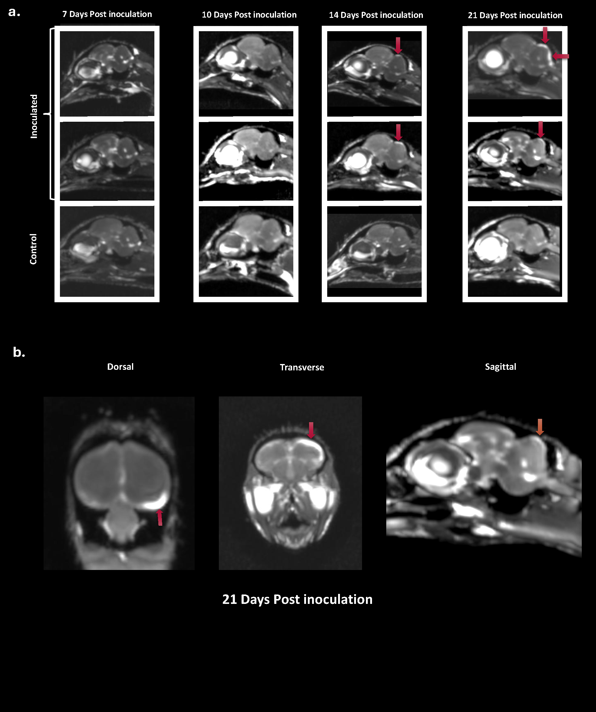
That collaboration with diagnostic imaging is one of the memorable parts of the story. In poultry medicine, radiology and advanced imaging are not always the first methods that come to mind when investigating infectious disease. Yet a conversation with Eberhard Ludewig from the Diagnostic Imaging Unit opened a new direction. His colleague Yasamin Vali was interested in birds, and soon the idea emerged to include magnetic resonance imaging, or MRI, in the study.
This turned out to be a major advantage. MRI allowed us to examine structural changes in the brains of experimentally infected birds in a way that complemented pathology and molecular testing. It helped us connect clinical disease, tissue lesions, and viral distribution with changes visible at the level of the whole organ.
The experimental infections were another key part of the study. Field observations are crucial because they show what happens under natural conditions, but they are rarely enough to establish whether a virus is truly involved in disease. Therefore, we performed in vivo infection studies in different avian species, including red-legged partridges, grey partridges, and chickens.
These experiments showed that the virus could spread systemically and reach neural tissues in different bird species. Importantly, in red-legged partridges, infection reproduced neurological disease and encephalitic lesions. This was a central finding of the work: the evidence was no longer based only on detecting a virus in diseased birds from the field. It was supported by experimental infection, pathology, imaging, molecular detection, and demonstration of viral replication.
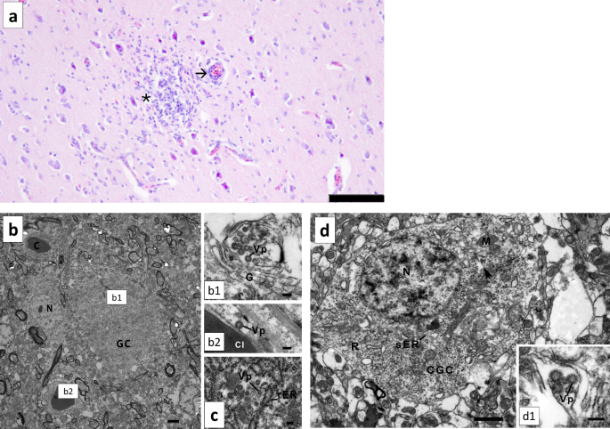
To build this evidence, we combined many techniques. Next-generation sequencing helped identify the virus. Phylogenetic analysis showed its relationship to other pegiviruses. RT-qPCR allowed us to detect and quantify viral RNA. Histopathology revealed the tissue lesions. Transmission electron microscopy provided ultrastructural information. RNAscope in situ hybridization showed where viral RNA was located within tissues. Immunohistochemistry, made possible by producing antibodies against part of the viral E2 protein, helped detect viral antigen. MRI added an imaging perspective on brain involvement. Finally, assays detecting viral replication supported the conclusion that the virus was not merely present, but active in infected tissues.
For us, this was one of the most satisfying aspects of the project: no single method carried the whole story. Instead, the evidence came from the convergence of many approaches.
The study also highlights the value of clinically driven research. The project did not begin as an abstract question about viral evolution or molecular mechanisms. It began with sick birds in the field. From that starting point, it expanded into a broader investigation with relevance for virology, pathology, comparative medicine, and host-virus interactions.
This is particularly important for pegiviruses. For many years, these viruses have been regarded as largely non-pathogenic. Recent reports in humans have raised questions about possible neurological involvement, but the field remains cautious, partly because detecting a common virus does not automatically mean that it causes disease. Our avian study contributes a different type of evidence. By combining natural outbreaks with experimental infections and multiple tissue-based methods, it provides strong support that an avian pegivirus can show neurotropism and be associated with encephalitis.
Of course, many questions remain. How does the virus reach the brain? Which cells support replication? Why are some avian species or individuals more affected than others? What determines whether infection remains subclinical or progresses to neurological disease? These are now important directions for future research.
Looking back, the project was a true collaborative exercise. It required field observations, clinical knowledge, pathology, molecular virology, experimental animal work, imaging, electron microscopy, and bioinformatics. It also required persistence through confusing moments, including multiple viral variants and technically demanding assay development.
For us, the paper represents more than the identification of a virus. It shows how a real clinical problem can lead to fundamental biological insight when different disciplines come together. It also reminds us that viruses considered harmless or poorly understood may still have surprises to reveal — if we follow the evidence carefully enough.
Our hope is that this work will stimulate further research into pegivirus biology, neurotropism, and pathogenesis, and encourage more attention to unexplained neurological diseases in animals. Sometimes, the most interesting discoveries begin not with a planned hypothesis, but with a question from the field: what is causing this disease?

Follow the Topic
-
Nature Communications
An open access, multidisciplinary journal dedicated to publishing high-quality research in all areas of the biological, health, physical, chemical and Earth sciences.

Related Collections
With Collections, you can get published faster and increase your visibility.
Neuroengineering
Publishing Model: Hybrid
Deadline: Oct 31, 2026
Women's Health
Publishing Model: Hybrid
Deadline: Ongoing