Intrapartum Ultrasound Image Analysis: Beginning from Standard Planes
Published in Bioengineering & Biotechnology, General & Internal Medicine, and Statistics
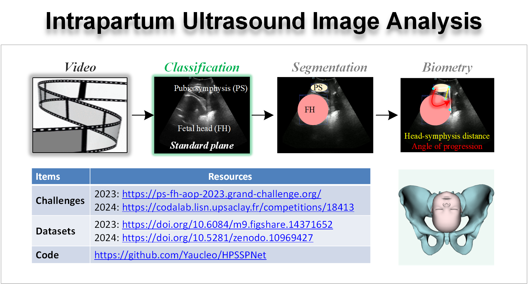
Ultrasound (US) imaging is extensively utilized in obstetric assessments due to its non-invasive nature, absence of radiation, affordability, and capability for real-time imaging. US imaging offers a clear advantage over traditional vaginal digital examinations by providing immediate, detailed visual insights into labor progression, including cervical dilation, fetal orientation, descent of the fetal head, and the rate of labor progression. These details are critical for physicians to make informed decisions about delivery management, potentially reducing neonatal mortality rates. The process of intrapartum US imaging is typically segmented into five steps: scanning, identification of the standard plane, observation of structures, measurement of parameters, and diagnosis. Among these, identifying the standard plane is vital, as it must include all critical structures necessary for accurate parameter measurements, directly impacting labor evaluation.
For instance, the angle of progression (AoP) is an essential metric in assessing the cephalopelvic fit during labor. Accurate AoP measurement requires clear visualization of the longitudinal sagittal plane of the pubic symphysis (PS) and the fetal head in US images. Real-time AoP monitoring is instrumental in predicting the mode of delivery, guiding clinical interventions, and minimizing risks to both mother and infant. Consequently, accurate identification of the pubic symphysis-fetal head standard plane (PSFHSP), defined as the intrapartum US image that distinctly portrays the fetal head and PS structures, is crucial for precise AoP measurements.
This study presents an innovative approach to automatically recognizing the PSFHSP in ultrasound imaging. By compressing the depth and modifying the residual modules of the model, we have enhanced its classification precision. Unlike traditional deep learning models, which typically require significant computational resources and substantial energy consumption, our proposed model is optimized for real-time classification on resource-constrained and low-energy edge devices. This makes it particularly well-suited for use with portable, handheld intrapartum ultrasound machines, offering a significant advancement in the field by facilitating efficient and reliable diagnostic assessments in various clinical environments.

The whole dataset used for the PSFHS challenge of MICCAI2023 (https://ps-fh-aop-2023.grand-challenge.org/) includes two parts: one is this PSFHS dataset (https://doi.org/https://doi.org/10.5281/zenodo.10969427) and another is from the JNU-IFM dataset (https://doi.org/10.6084/m9.figshare.14371652). These images can also be used for the Intrapartum Ultrasound Grand Challenge (IUGC) 2024 of MICCAI 2024 (https://codalab.lisn.upsaclay.fr/competitions/18413). For transparency and reproducibility, the source code of our model has been made publicly accessible at https://github.com/Yaucleo/HPSSPNet.
Finally, we are delighted to share our work with the scientific community and domain experts in the prestigious journal, Medical & Biological Engineering & Computing. We sincerely hope that this resource can provide valuable research groundwork and further insights for the community.
Reference
Fernandez-Turienzo C, Sandall J. Delivering high-quality childbirth care. Nat Med. 2024;30(2):348-349. doi:10.1038/s41591-024-02812-2
Zhou M, Wang C, Lu Y, et al. The segmentation effect of style transfer on fetal head ultrasound image: a study of multi-source data. Med Biol Eng Comput. 2023;61(5):1017-1031. doi:10.1007/s11517-022-02747-1
Ou, Z., Bai, J., Chen, Z., Lu, Y., Wang, H., Long, S., Chen, G., RTSeg-Net: A Lightweight Network for Real-time Segmentation of Fetal Head and Pubic Symphysis from Intrapartum Ultrasound Images. Computers in biology and medicine, 2024; 108501. doi:10.1016/j.compbiomed.2024.108501
Qiu, R., Zhou M, Bai, J., Lu, Y., Wang, H. PSFHSP-Net: An Efficient Lightweight Network for Identifying Pubic Symphysis-Fetal Head Standard Plane from Intrapartum Ultrasound Images. Med Biol Eng Comput. 2024. https://doi.org/10.1007/s11517-024-03111-1
Chen, Z. Ou, Y. Lu, J. Bai, Direction-guided and multi-scale feature screening for fetal head–pubic symphysis segmentation and angle of progression calculation, Expert Systems with Applications, 245 (2024) 123096. doi:10.1016/j.eswa.2023.123096
Lu Y, Zhou M, Zhi D, et al. The JNU-IFM dataset for segmenting pubic symphysis-fetal head [published correction appears in Data Brief. 2022 Apr 01;42:108128]. Data Brief. 2022;41:107904. Published 2022 Feb 2. doi:10.1016/j.dib.2022.107904
Lu Y, Zhi D, Zhou M, et al. Multitask Deep Neural Network for the Fully Automatic Measurement of the Angle of Progression. Comput Math Methods Med. 2022;2022:5192338. Published 2022 Sep 2. doi:10.1155/2022/5192338
Bai J, Sun Z, Yu S, et al. A framework for computing angle of progression from transperineal ultrasound images for evaluating fetal head descent using a novel double branch network. Front Physiol. 2022;13:940150. Published 2022 Dec 2. doi:10.3389/fphys.2022.940150
Chen G, Bai J, Ou Z, Lu Y, Wang H. PSFHS: Intrapartum ultrasound image dataset for AI-based segmentation of pubic symphysis and fetal head. Scientific Data. 2024. doi:10.1038/s41597-024-03266-4
Chen, Y. Lu, S, Long, V, et al. Fetal Head and Pubic Symphysis Segmentation in Intrapartum Ultrasound Image Using a Dual-Path Boundary-Guided Residual Network. IEEE Journal of Biomedical and Health Informatics. 2024
Follow the Topic
-
Medical & Biological Engineering & Computing

This journal covers the entire spectrum of biomedical and clinical engineering. The journal aims to present exciting and vital experimental and theoretical developments in biomedical science and technology and to report on advances in computer-based methodologies in these multidisciplinary subjects.


Please sign in or register for FREE
If you are a registered user on Research Communities by Springer Nature, please sign in