JMCR: Clinical Reasoning from Case Reports
Published in Biomedical Research
Explore the Research
JavaScript is disabled
The Clinical Problem
An 18-year-old man presented with six months of nocturnal pain in the proximal right thigh. Nonsteroidal anti-inflammatory medication relieved the pain.
Examination showed normal hip movement with mild quadriceps atrophy. Routine laboratory studies—including complete blood count, erythrocyte sedimentation rate, alkaline phosphatase, and lactate dehydrogenase—were normal.
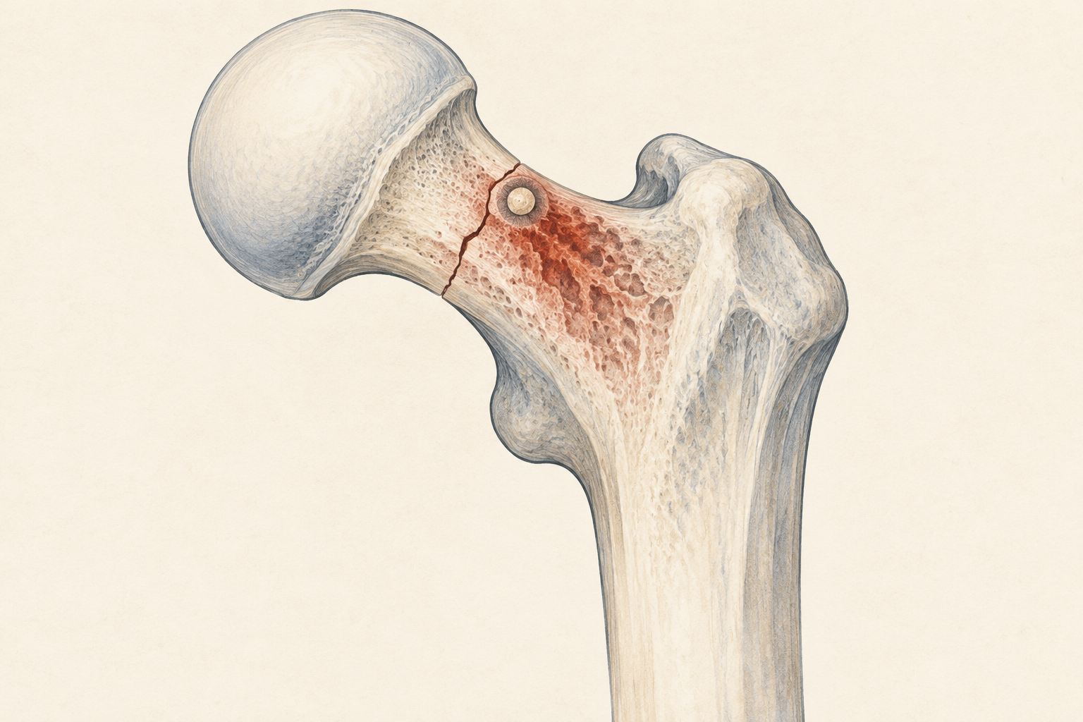
Radiography and CT demonstrated two small, well-defined nidus-like lesions in the femoral neck, including a subperiosteal lytic lesion with central ossification and surrounding reactive bone.
The symptoms and imaging appeared to point toward osteoid osteoma.
But there was an early discordant feature: the apparent multifocality.
Clinical Reasoning: Step 1
Why was osteoid osteoma initially convincing?
The patient had:
- young age;
- nocturnal focal pain;
- relief with an NSAID;
- a small, well-defined lytic lesion;
- central mineralization; and
- surrounding reactive sclerosis.
These are familiar features of osteoid osteoma.
A core biopsy was performed, and the sampled tissue did not contain malignant spindle cells or malignant osteoid. Following tumor-board discussion, the lesions were classified as multifocal osteoid osteoma.
Radiofrequency ablation was performed, and the pain resolved.
Pause and consider
Does an initial treatment response confirm the diagnosis?
No. Pain relief following local ablation is compatible with successful treatment of osteoid osteoma, but it is not histological proof and does not guarantee that the entire biological process was sampled or treated.
Clinical Reasoning: Step 2
Which initial feature deserved greater scrutiny?
Two discrete nidus-like abnormalities were identified.
Multifocal osteoid osteoma is unusual. The presence of more than one lesion should therefore have widened the differential diagnosis and increased concern about whether a limited biopsy was representative.
Important alternatives for a cortical lytic lesion include:
- osteoid osteoma;
- osteoblastoma;
- intracortical osteosarcoma;
- stress fracture;
- intracortical infection;
- fibrous dysplasia;
- transcortical chondroma; and
- reactive periosteal lesions.
Clinical reasoning point
A rare presentation of a common benign diagnosis may be correct—but it should also prompt reconsideration of a rarer mimic.
Clinical Reasoning: Step 3
The return of pain changes the interpretation
The patient remained pain-free for approximately one year. Nocturnal pain then returned, and radiographs showed incomplete ossification of the lesion.
He declined surgery and continued NSAID treatment.
At this stage, the earlier diagnosis should no longer have been treated as settled. Persistent or recurrent symptoms after technically directed treatment may reflect incomplete ablation, recurrence, an alternative pain source, or an incorrect original diagnosis.
The key is not that every recurrence means malignancy. It is that recurrence creates new evidence that must be reconciled with the original conclusion.
Clinical Reasoning: Step 4
The pathological fracture becomes the decisive warning
Eighteen months later, the patient presented with a femoral-neck fracture.
MRI now demonstrated a heterogeneous femoral-neck mass with soft-tissue extension and surrounding edema.
These features could no longer be reconciled adequately with a small, uncomplicated osteoid osteoma.
Repeat core biopsy showed malignant spindle cells producing osteoid.
Diagnostic pivot
The finding of malignant osteoid established osteosarcoma. The lesion’s predominantly cortical location supported the classification of intracortical osteosarcoma.
Clinical Reasoning: Step 5
Why could the first biopsy have been negative?
The original specimen did not contain malignant spindle cells or malignant bone.
The authors considered at least two possible explanations:
- Sampling error: A small core may not have captured the malignant component of a heterogeneous lesion.
- Subtle early histology: A low-grade or limited early component may have been difficult to recognize.
The published information cannot determine conclusively which explanation applies.
Clinical pearl
A negative biopsy is reassuring only to the extent that the specimen adequately represents the radiological target and agrees with the subsequent clinical course.
What Was the Final Diagnosis?
Intracortical conventional osteosarcoma of the femoral neck, initially interpreted and treated as multifocal osteoid osteoma.
Staging CT and bone scintigraphy found no distant metastases.
The patient underwent wide resection with frozen-section assessment of the margins and reconstruction using an allograft–prosthesis composite. Histopathology confirmed conventional osteosarcoma.
He subsequently completed adjuvant methotrexate, doxorubicin, and cisplatin chemotherapy. At four years, he was alive and pain-free, without reported local recurrence or distant metastasis.
This individual outcome does not establish the comparative effectiveness of the reconstructive or chemotherapy strategy.
Differential Diagnosis at the Critical Decision Point
1. Osteoid osteoma
The age, nocturnal NSAID-responsive pain, nidus-like appearance, and reactive sclerosis strongly supported this diagnosis initially. Multifocality, recurrent pain, incomplete ossification, and later pathological fracture became increasingly discordant.
2. Osteoblastoma
Osteoblastoma may resemble osteoid osteoma histologically and radiologically, particularly when lesions are larger. The ultimate identification of malignant spindle cells producing osteoid excluded a benign osteoblastoma.
3. Intracortical osteosarcoma
Its rarity made it initially less likely, but cortical location, diagnostic discordance, progressive behavior, soft-tissue extension, pathological fracture, and malignant osteoid ultimately supported the diagnosis.
4. Stress fracture
A femoral-neck stress injury can cause pain and reactive bone change. The two discrete nidus-like lesions and later malignant histopathology argued against it.
5. Intracortical infection
An intracortical abscess can produce a small lytic lesion with reactive sclerosis. Normal inflammatory studies did not exclude infection, but the subsequent biopsy demonstrated malignancy.
6. Fibrous dysplasia or another benign fibro-osseous lesion
These may affect the proximal femur but did not explain the final high-grade spindle-cell proliferation with malignant osteoid.
Why This Case Matters
The original diagnosis was not irrational. It matched the patient’s symptoms and much of the imaging appearance.
The error arose because later evidence no longer fit the original model.
Clinical reasoning requires more than reaching a plausible diagnosis. It requires continued testing of that diagnosis against the patient’s subsequent trajectory.
In this case, multifocality was the early warning; recurrent pain was the second warning; incomplete healing and pathological fracture made diagnostic reopening unavoidable.
Clinical Pearls
- NSAID-responsive nocturnal pain strongly suggests osteoid osteoma but is not disease-specific.
- Multifocal osteoid osteoma is unusual and should prompt careful reconsideration of mimics.
- A limited biopsy may not represent a heterogeneous bone lesion.
- Clinical, radiological, and pathological findings should agree before ablation.
- Temporary improvement after treatment does not retrospectively prove the diagnosis.
- Recurrent pain or incomplete ossification warrants renewed imaging review.
- A pathological fracture is a major change in biological behavior.
- Malignant osteoid produced by malignant cells is the defining pathological feature of osteosarcoma.
Potential Pitfalls
Pitfall 1: Allowing classic symptoms to override an atypical feature
Better approach: Recognize the osteoid-osteoma pattern while explicitly addressing why two nidus-like lesions are present.
Pitfall 2: Treating the initial biopsy as definitive despite possible sampling limitations
Better approach: Confirm that the specimen represents the most suspicious radiological component and reconsider biopsy when clinicoradiological discordance develops.
Pitfall 3: Using pain relief after ablation as diagnostic confirmation
Better approach: Regard treatment response as follow-up information, not a substitute for adequate pathological correlation.
Pitfall 4: Explaining recurrent symptoms as routine recurrence without reassessment
Better approach: Repeat imaging and reconsider the full differential when the lesion does not heal or behaves more aggressively than expected.
Clinical Timeline
| Time | Clinical development |
|---|---|
| Initial presentation | Six months of nocturnal proximal-thigh pain relieved by NSAIDs |
| Initial imaging | Two small nidus-like femoral-neck lesions with reactive bone |
| Initial biopsy | No malignant spindle cells or malignant osteoid identified |
| Initial management | Tumor board favors multifocal osteoid osteoma; radiofrequency ablation performed |
| One year | Pain returns; incomplete lesion ossification is seen |
| Approximately 18 months later | Femoral-neck pathological fracture and heterogeneous mass with soft-tissue extension |
| Diagnostic pivot | Repeat biopsy demonstrates malignant spindle cells producing osteoid |
| Definitive treatment | Wide resection, allograft–prosthesis reconstruction, and adjuvant MAP chemotherapy |
| Four-year follow-up | Alive and pain-free without reported recurrence or distant metastasis |
Test Your Clinical Reasoning
Question 1
Which feature was least typical of an ordinary solitary osteoid osteoma at the initial presentation?
A. The patient’s age
B. Nocturnal pain
C. Pain relief with NSAIDs
D. Two separate nidus-like lesions
E. Surrounding reactive bone
Answer: D. Two separate nidus-like lesions
Rationale: The other findings are characteristic of osteoid osteoma. Apparent multifocality is unusual and should broaden the differential diagnosis.
Question 2
Which later finding most clearly required abandonment of the original benign diagnostic framework?
A. Normal alkaline phosphatase
B. Temporary pain relief after ablation
C. Pathological femoral-neck fracture with an enlarging mass
D. Mild quadriceps atrophy
E. Normal inflammatory markers
Answer: C. Pathological femoral-neck fracture with an enlarging mass
Rationale: A pathological fracture and progressive mass demonstrate biological behavior incompatible with a successfully treated, uncomplicated osteoid osteoma.
Question 3
What finding established the diagnosis of osteosarcoma?
A. NSAID-responsive pain
B. Reactive cortical sclerosis
C. Malignant spindle cells producing osteoid
D. A femoral-neck location
E. Absence of distant metastases
Answer: C. Malignant spindle cells producing osteoid
Rationale: Malignant osteoid produced by malignant tumor cells is the defining pathological feature.
Question 4
How should the original negative biopsy be interpreted?
A. It permanently excluded malignancy
B. It proved that malignant transformation later occurred
C. It may have been limited by sampling error or subtle early histology
D. It established osteoblastoma
E. It made further biopsy unnecessary
Answer: C. It may have been limited by sampling error or subtle early histology
Rationale: The report cannot determine whether the first specimen missed the malignant component or contained changes too limited for recognition.
Question 5
What is the broader diagnostic lesson?
A. Every osteoid osteoma should undergo wide resection
B. Radiofrequency ablation causes osteosarcoma
C. A diagnosis should be reopened when the subsequent course becomes discordant
D. Normal laboratory tests exclude bone malignancy
E. Temporary treatment response establishes histology
Answer: C. A diagnosis should be reopened when the subsequent course becomes discordant
Rationale: Multifocality, recurrent symptoms, incomplete healing, and pathological fracture provided accumulating evidence against the original diagnosis.
Clinical Take-Home Message
A diagnosis of osteoid osteoma may appear convincing when a young patient has nocturnal NSAID-responsive pain and a small nidus-like cortical lesion.
However, multifocality, recurrent pain after treatment, incomplete ossification, lesion progression, or pathological fracture should prompt renewed imaging review and repeat tissue assessment. A negative initial biopsy does not permanently exclude malignancy when sampling may have been limited and the later clinical course no longer fits.
More broadly, this case reminds us that diagnostic accuracy depends not only on the initial evidence but also on whether the diagnosis continues to explain what happens next.
Journal of Medical Case Reports is the world’s first international, PubMed-listed medical journal devoted to publishing case reports from all medical disciplines and will consider any original case report that expands the field of general medical knowledge, and original research relating to case reports. The journal is open access and strongly endorses the CARE guidelines for case reports, requiring authors to submit populated CARE checklists with submissions to improve transparency in reporting.

Richard Alan Rison is the Editor-in-Chief of Journal of Medical Case reports. He is also an associate neurology editor (editorial board) for BMC Neurology, and the former lead editor for case reports of BMC Research Notes (currently on the editorial board). His scholarly work focuses on medical case reporting, reporting standards, and editorial methodology. Dr. Rison participated in the development and dissemination of the CARE guidelines for clinical case reporting and has authored numerous publications addressing both neurological disorders and the role of case reports in advancing medical knowledge. Dr. Rison practices general neurology and served as the founding medical director of the PIH Health Hospital-Whittier Stroke Program and the PIH Health Hospital-Whittier Non-Invasive Vascular Laboratory, is a clinical assistant professor of neurology at the University of Southern California Keck School of Medicine and Los Angeles County Medical Center, and is a Fellow of the American Academy of Neurology, the American Neurological Association, and the American Association of Neuromuscular and Electrodiagnostic Medicine. Dr Rison is board-certified by the American Board of Psychiatry and Neurology in neurology and vascular neurology, and neurocritical care and neuroimaging by the United Council of Neurologic Subspecialties. He is also board-certified by the American Board of Electrodiagnostic Medicine in electrodiagnostic medicine. Dr. Rison is a former president of the Los Angeles Neurological Society.
Follow the Topic
-
Journal of Medical Case Reports
This journal will consider any original case report that expands the field of general medical knowledge, and original research relating to case reports.