JMCR: Highlight
Published in Biomedical Research
Explore the Research
Coral reef aorta in a child: a case report
Severe hypertension in a child demands urgent investigation. When it is accompanied by organ infarction and complex vascular abnormalities, the diagnostic challenge becomes even greater.
Our recent case report describes a 12-year-old Uyghur girl who presented with headache and exceptionally high blood pressure, reaching 225/158 mmHg. Imaging also revealed renal and splenic infarctions, suggesting significant systemic vascular disease. Given her age and the pattern of arterial involvement, polyarteritis nodosa was initially considered the most likely diagnosis.
However, the full vascular picture told a different story.
An unexpected diagnosis
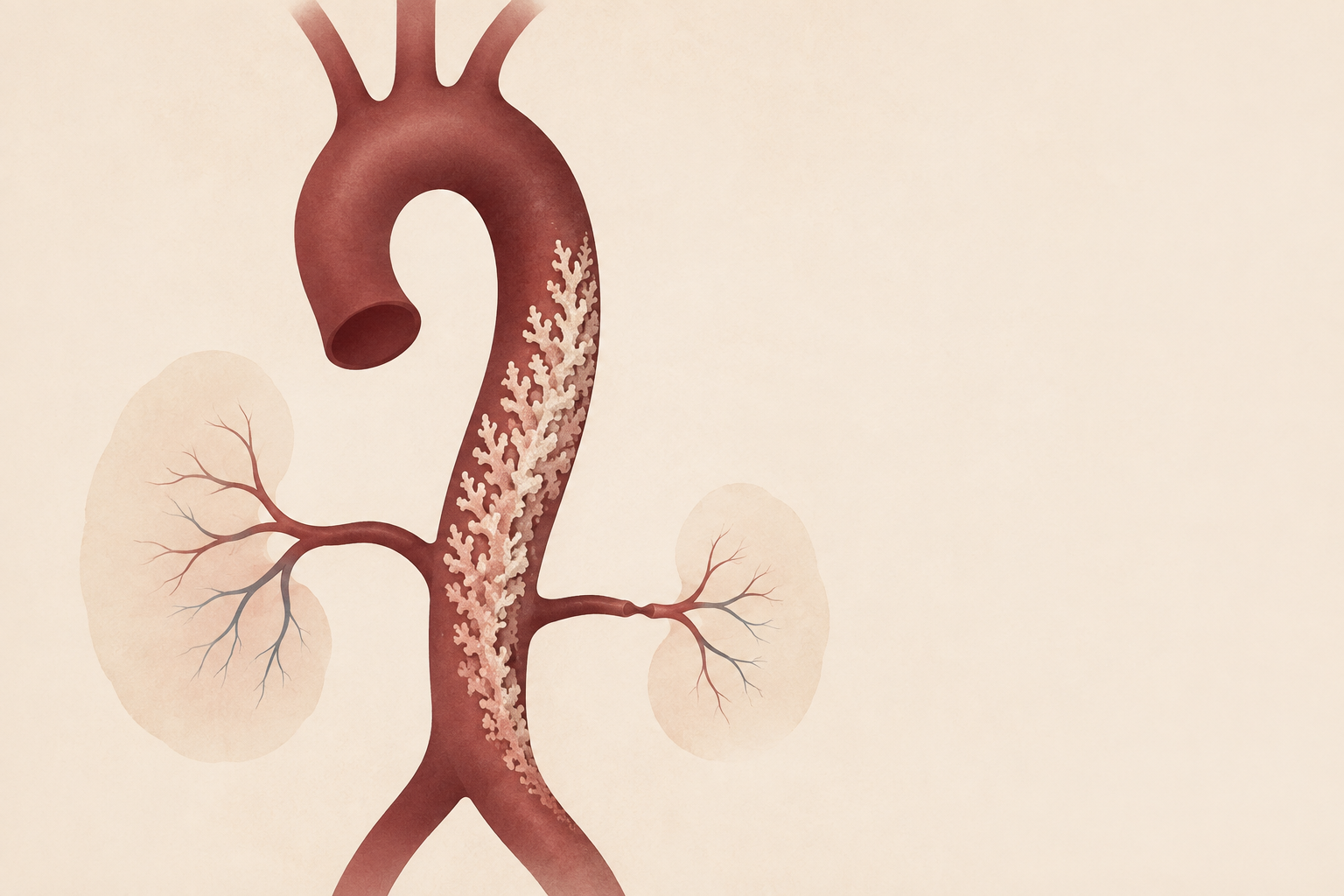
Whole-aortic computed tomography angiography demonstrated irregular, densely calcified plaques projecting into the lumen of the thoracic and abdominal aorta. Their striking, rock-like appearance resembled a coral reef. Renal artery stenosis and occlusion of additional aortic branch vessels were also present.
These findings led to the diagnosis of coral reef aorta, a rare form of obstructive aortic disease characterized by heavily calcified intraluminal lesions. The condition is reported predominantly in adults and is exceptionally uncommon in children.
The resemblance to systemic vasculitis created an important diagnostic pitfall. Both conditions may produce severe hypertension, arterial stenosis, organ ischemia and involvement of multiple vascular territories. In this case, however, the distinctive calcified morphology and distribution of the aortic lesions supported coral reef aorta rather than primary inflammatory vasculitis.
Why imaging the entire aorta mattered
A central lesson from this case is the value of early, comprehensive vascular imaging.
In a child with refractory hypertension, investigation often focuses on the renal arteries and other common secondary causes. A limited examination, however, may fail to reveal disease extending throughout the thoracic and abdominal aorta.
Whole-aortic angiography allowed us to recognize the characteristic calcified plaques, determine the extent of branch-vessel involvement and select an appropriate intervention. It also helped distinguish a structural obstructive disorder from vasculitis, two diagnoses that require markedly different treatment strategies.
Restoring renal blood flow
Because renal artery stenosis was considered a major contributor to the patient’s hypertension, balloon angioplasty was performed alongside antihypertensive therapy. This approach achieved sustained blood-pressure control. At 18 months of follow-up, the patient had experienced no recurrence of hypertension.
The favorable outcome illustrates the importance of timely recognition and targeted treatment. Persistent severe hypertension during childhood can rapidly produce irreversible injury to the kidneys, heart, brain and other organs. When renovascular obstruction is identified, restoring blood flow may address an important underlying driver rather than relying solely on medical blood-pressure control.
A rare disease with a practical message
The rarity of coral reef aorta in childhood makes it easy to overlook. Nevertheless, this case offers a broadly applicable clinical message: unusual diagnoses should remain under consideration when the imaging pattern does not fully support the initial explanation.
For children presenting with unexplained refractory hypertension—particularly when accompanied by organ ischemia or evidence of multivessel disease—clinicians should consider imaging the entire aorta and its major branches. Careful attention to the morphology of vascular lesions may be just as important as identifying where stenosis or occlusion has occurred.
Recognizing the “coral reef” appearance in this patient changed the diagnosis, guided treatment and helped prevent further end-organ injury. We hope this case increases awareness of a rare but consequential vascular disorder and encourages comprehensive evaluation when childhood hypertension follows an unexpected course.
Journal of Medical Case Reports is the world’s first international, PubMed-listed, medical journal devoted to publishing case reports from all medical disciplines and will consider any original case report that expands the field of general medical knowledge, and original research relating to case reports. The journal is open access, and strongly endorses the CARE guidelines for case reports, requiring authors to submit populated CARE checklists with submissions to improve transparency in reporting.

Richard Alan Rison is the Editor-in-Chief of Journal of Medical Case reports. He is also an associate neurology editor (editorial board) for BMC Neurology, and the former lead editor for case reports of BMC Research Notes (currently on the editorial board). His scholarly work focuses on medical case reporting, reporting standards, and editorial methodology. Dr. Rison participated in the development and dissemination of the CARE guidelines for clinical case reporting and has authored numerous publications addressing both neurological disorders and the role of case reports in advancing medical knowledge. Dr. Rison practices general neurology and served as the founding medical director of the PIH Health Hospital-Whittier Stroke Program and the PIH Health Hospital-Whittier Non-Invasive Vascular Laboratory, is a clinical assistant professor of neurology at the University of Southern California Keck School of Medicine and Los Angeles County Medical Center, and is a Fellow of the American Academy of Neurology, the American Neurological Association, and the American Association of Neuromuscular and Electrodiagnostic Medicine. Dr Rison is board-certified by the American Board of Psychiatry and Neurology in neurology and vascular neurology, and neurocritical care and neuroimaging by the United Council of Neurologic Subspecialties. He is also board-certified by the American Board of Electrodiagnostic Medicine in electrodiagnostic medicine. Dr. Rison is a former president of the Los Angeles Neurological Society.
Follow the Topic
-
Journal of Medical Case Reports
This journal will consider any original case report that expands the field of general medical knowledge, and original research relating to case reports.