Mapping dental stem-cell research: looking beyond publication counts

Dental stem-cell research has expanded substantially over the past 25 years, but publication volume alone does not necessarily show how different subtypes are discussed in relation to translation-oriented research.
Mapping dental stem-cell research: looking beyond publication counts
Like

Share this post

Choose a social network to share with, or copy the URL to share elsewhere

This is a representation of how your post may appear on social media. The actual post will vary between social networks

Explore the Research

Springer Berlin Heidelberg
Springer Berlin Heidelberg Springer Berlin Heidelberg

Bibliometric and science-mapping analysis of translation-oriented signals across six dental stem-cell subtypes: a Web of Science study (2000–2025) - Oral and Maxillofacial Surgery

Objectives To benchmark publication growth, conceptual structure, and translation-oriented signals across six dental stem-cell (DSC) subtypes from 2000 to 2025. Methods Web of Science Core Collection (SCI-E/SSCI/ESCI) was searched (2000–2025) for DPSC, PDLSC, SHED, SCAP, DFSC, and GMSC records. After cross-subtype deduplication, corpus-level indicators and networks were computed on the deduplicated corpus (N = 13,327) using fractional assignment to avoid multi-subtype inflation. Subtype-level time trends were computed within each subtype export (full counting). Translation-oriented signals (biomaterials/scaffolds, in vivo models, EVs/exosomes, clinical-trial mentions) were estimated from a stratified random sample of 500 records per subtype (fixed seed 20251216) using rule-based dictionaries with manual verification of sampled classifications. Results Output increased sharply after 2010 and spanned 1,462 journals. DPSC and PDLSC dominated publication volume, while SHED, SCAP, DFSC, and GMSC showed later growth. Science mapping resolved four recurring themes (regeneration applications; biomaterials/differentiation; angiogenesis/VEGF; immunomodulation/EVs). Across subtypes, biomaterials/scaffold and in vivo-model signals were frequent, EV/exosome signals rose after 2015, and clinical-trial mentions remained uncommon; SHED and GMSC showed comparatively higher EV/exosome and trial-mention proportions. Conclusions This subtype-resolved, overlap-aware evidence map provides a basis for comparative benchmarking and highlights where translational emphasis is increasing versus where clinical evidence remains sparse. Clinical relevance These findings may help inform model selection, biomaterials strategy design, and prioritization of evidence gaps in regenerative dentistry.

When I started working with dental stem cells and regenerative dentistry, I often noticed that some cell types appeared much more frequently in the literature than others. Dental pulp stem cells and periodontal ligament stem cells were widely represented, while other subtypes, such as SHED, SCAP, DFSC, and GMSC, seemed to follow different research trajectories.

This made me wonder: are the most frequently published dental stem-cell subtypes also the ones more often associated with translation-oriented language in the literature?

That question became the starting point for my recently published article in Oral and Maxillofacial Surgery. Instead of looking only at publication volume, I wanted to compare six dental stem-cell subtypes within the same analytical framework and examine how the literature has developed over 25 years.

As a single-author study, the process also required careful checking of terminology, overlap, and interpretation at each step.

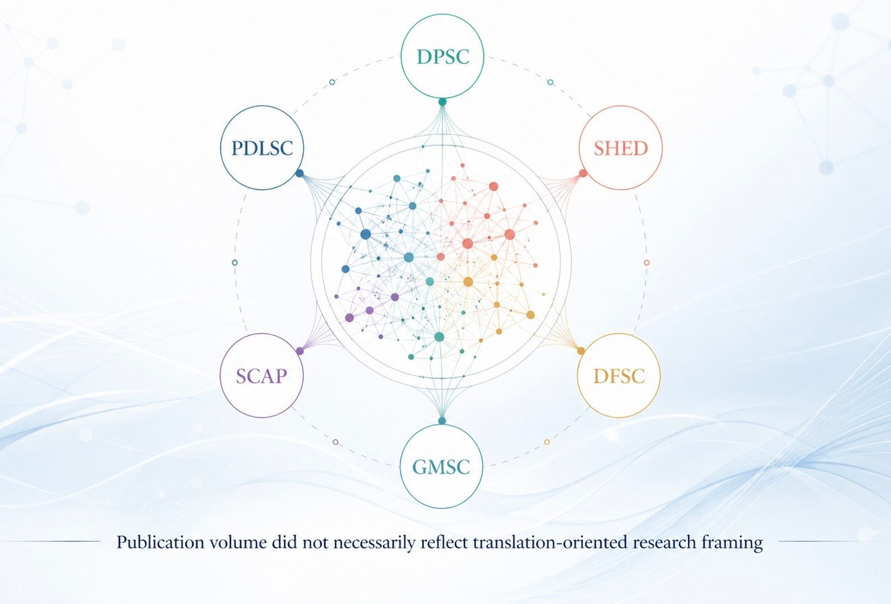
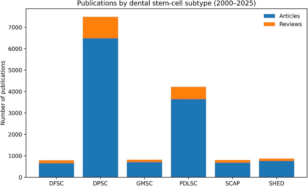
The study used Web of Science-indexed records from 2000 to 2025 and focused on six major dental stem-cell subtypes: dental pulp stem cells, periodontal ligament stem cells, stem cells from human exfoliated deciduous teeth, stem cells from the apical papilla, dental follicle stem cells, and gingival mesenchymal stromal/stem cells.

One challenge was that these literatures overlap. A single paper may mention more than one dental stem-cell subtype, which can inflate simple publication counts. To reduce this issue, the study used a deduplicated corpus and an overlap-aware fractional counting approach for corpus-level comparisons.

I chose Web of Science Core Collection as the single database because the study depended on a consistent citation backbone for document co-citation and science-mapping analyses. Combining databases such as Web of Science and Scopus can broaden coverage, but it can also introduce differences in indexing, duplicate records, citation formats, and network structures. For this reason, I treated the Web of Science-only design as a methodological choice for internal consistency, while also recognizing it as a limitation rather than complete coverage of the field.

I also wanted to be careful with interpretation. The study did not assess therapeutic efficacy, clinical readiness, or regulatory maturity. Instead, it examined selected translation-oriented signals in the literature, including terms related to biomaterials/scaffolds, in vivo models, extracellular vesicles/exosomes, and clinical-trial-related language. These signals should be understood as indicators of research framing, not as evidence of clinical effectiveness.

Within these limits, the analysis suggested that publication volume and translation-oriented framing were not always aligned. Earlier-established subtypes continued to dominate total publication output, while some later-emerging subtypes showed proportionally higher representation of selected translation-oriented signals.

For me, the value of this work is not in ranking one subtype above another. Rather, it provides a structured map of how the field is being discussed and where evidence gaps may remain. I hope it can help researchers interested in regenerative dentistry, dental stem cells, biomaterials, extracellular vesicles, and translational tissue engineering think more carefully about subtype-specific research emphasis.

Published article:
Zahedah R. Bibliometric and science-mapping analysis of translation-oriented signals across six dental stem-cell subtypes: a Web of Science study (2000–2025). Oral and Maxillofacial Surgery. 2026;30:79.

https://doi.org/10.1007/s10006-026-01564-2

Please sign in or register for FREE

If you are a registered user on Research Communities by Springer Nature, please sign in

Follow the Topic

Regenerative Medicine and Tissue Engineering
Life Sciences > Biological Sciences > Biotechnology > Regenerative Medicine and Tissue Engineering
Oral and Maxillofacial Surgery
Life Sciences > Health Sciences > Surgery > Oral and Maxillofacial Surgery
Dentistry
Life Sciences > Health Sciences > Clinical Medicine > Dentistry
Stem Cell Biology
Life Sciences > Health Sciences > Biomedical Research > Stem Cell Biology
Translational Research
Life Sciences > Health Sciences > Biomedical Research > Translational Research
Continuing professional development in dentistry
Life Sciences > Health Sciences > Clinical Medicine > Dentistry > Dental Education > Continuing professional development in dentistry

Ask the Editor - Oral and Maxillofacial Surgery

Got a question for the editor about Malocclusions or misalignment of the teeth? Ask it here!

Continue reading announcement

Related Collections

With Collections, you can get published faster and increase your visibility.

Current State and Future Opportunities for 3D Printing/Additive Manufacturing in Oral and Maxillofacial Surgery: from Basic Science to Clinical Applications

Three-dimensional (3D) printing, also known as additive manufacturing, has emerged as a transformative technology in oral and maxillofacial surgery. It offers unprecedented precision and customization in the fabrication of patient-specific anatomical models, surgical guides, and implants. This not only enhances preoperative planning and intraoperative accuracy but also significantly reduces surgical time and improves postoperative outcomes, as surgeons can now visualize complex craniofacial structures in tangible form, simulate procedures, and tailor interventions to the unique anatomical and functional needs of each patient. It minimizes risks and optimizes aesthetic and functional results, especially in cases involving congenital deformities, trauma reconstruction, oncologic resections, and orthognathic surgery, where traditional methods often fall short in achieving ideal fit and alignment. The integration of biocompatible materials and advances in digital imaging and computer-aided design (CAD) further expand the scope of 3D printing applications, enabling the production of implants that conform precisely to bone contours and support tissue regeneration. As this technology continues to evolve, with ongoing research into bio-printing and the use of stem cells and growth factors, the potential for creating living tissue constructs and fully functional bone grafts becomes increasingly feasible, promising a future where personalized, regenerative solutions become standard practice in maxillofacial care. Although challenges remain in terms of regulatory approval, cost-effectiveness, and widespread clinical adoption, the growing body of evidence supporting its efficacy and safety underscores the importance of continually educating practitioners on the latest developments in the subject to enable informed decision-making and ultimately, improved patient outcomes.

Publishing Model: Hybrid

Deadline: Ongoing