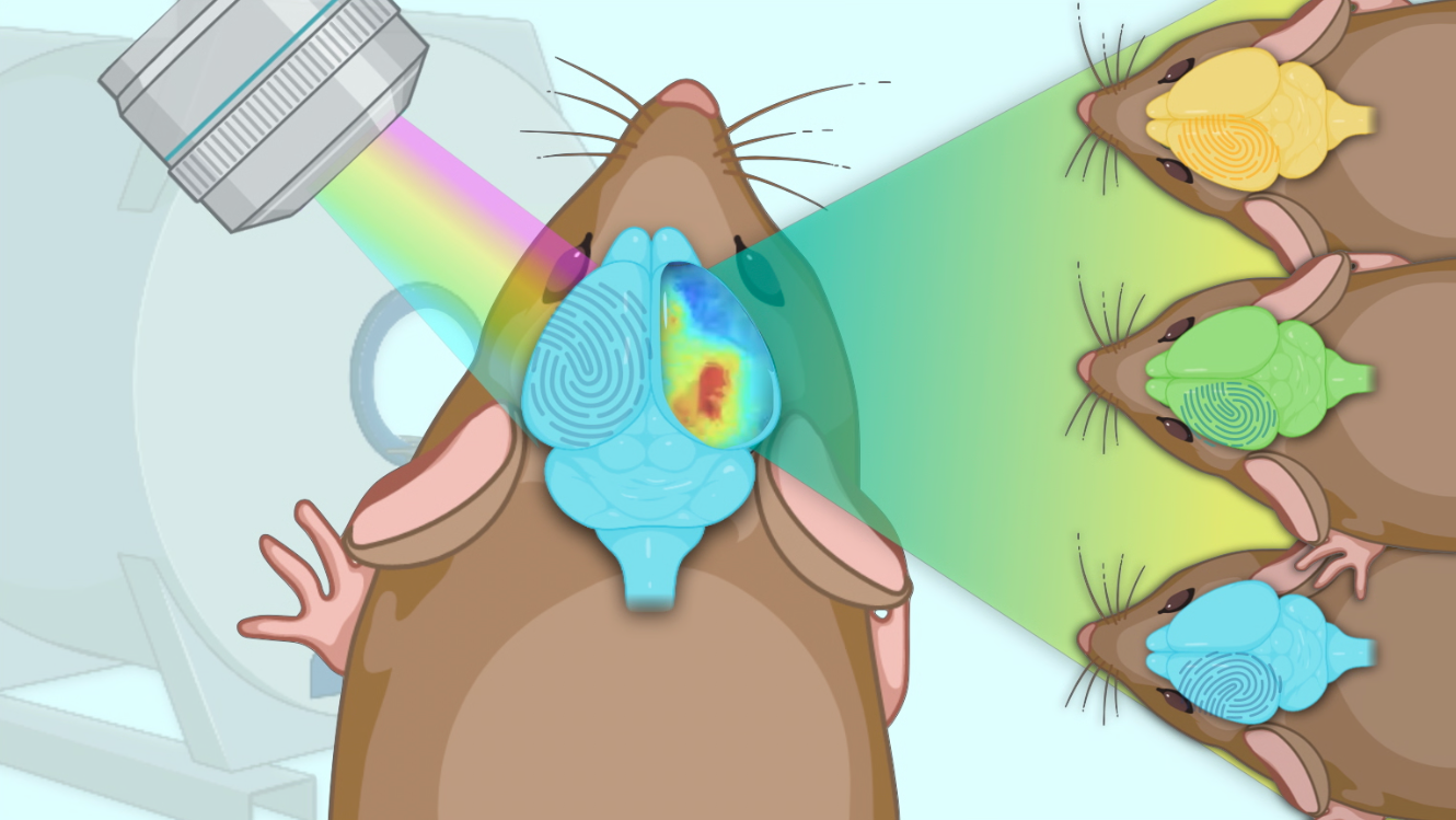
Multimodal identification of the mouse brain using simultaneous Ca2+ imaging and fMRI
Published in Physics, Protocols & Methods, and General & Internal Medicine

Studying individuals, not averages.
No two individuals are perfectly alike—even in the case of identical twins. Nevertheless, standard scientific and medical approaches often categorize people into groups. There is a growing recognition in clinical and cognitive neuroscience1,2 that a more precise approach is needed, echoing recent advancements in oncology3 and cardiovascular disease4.
One approach to study individual differences in blood-oxygen-level-dependent (BOLD) functional magnetic resonance imaging (fMRI) is to compare an individual’s data to others in a process called connectome-based identification (ID)5. This procedure identifies subjects from a pool using their connectome (a summary of inter-regional BOLD signal synchronies or connectivity strengths). High rates of ID are typical in human populations—so much so, that it is common to think of an individual’s connectome as akin to a ‘fingerprint'5-7.
Understanding across spatial scales.
However, there are many unanswered questions related to an individual’s brain connectome fingerprint. Is the individual uniqueness due to true underlying brain differences, or is it due to something else—perhaps due to the nature of BOLD-fMRI itself? For all of its strengths, BOLD-fMRI suffers from low spatiotemporal resolution and specificity as well as a propensity for noise and spurious signal corruption. To help address this question, we can use animal models in which fMRI, alongside complementary but more invasive contrasts, can be accessed.
To better appreciate the relationships between the BOLD signal and underlying cellular activity, we imaged mice using simultaneous wide-field fluorescence calcium imaging and BOLD-fMRI8. The use of wide-field fluorescence calcium imaging allowed us to target the recording of specific cell types, including excitatory neurons, inhibitory neurons, and glial cells. Beyond helping us address the cellular aspects of individual differences in the BOLD signal, the use of multiple imaging modalities measured simultaneously allowed us to compare results across multiple spatiotemporal scales, affording a more complete picture of the dynamics associated with neural activity.
What we found.
We found that we could significantly ID individual mice using both WF-Ca2+ data and BOLD-fMRI data9. In addition, we found we could successfully predict, in both WF-Ca2+ data and BOLD-fMRI data, the connectivity signatures associated with the cell-type that was labelled (i.e., mice expressing a fluorescent label in excitatory neurons tended to look more like other mice expressing a fluorescent label in excitatory neurons). Interestingly, the ID rates obtained using WF-Ca2+ data were consistently statistically significantly higher than the ID rates obtained using BOLD-fMRI data. We also examined whether we could ID mice across modality (using the WF-Ca2+ connectome to ID the BOLD-fMRI connectome); we found this was not successful.
Why this matters.
Our finding of high ID rates in the WF-Ca2+ imaging data supports the notion that individual differences observed in BOLD-fMRI are due to cellular differences. Nevertheless, BOLD-fMRI ID rates were still quite high (albeit not as high as those using the WF-Ca2+ data). The fact that cross-modal ID was not possible suggests that each imaging modality offers complementary information about individual-specific connectivity signatures and reinforces that much exciting work remains to be conducted linking data across spatiotemporal scales. More generally, the work performed here supports the continued investigation of individual differences in connectomes. It also suggests that focusing on individual-based approaches in animal studies—instead of only focusing on groups—is a valid approach that can potentially teach us much about neurobiology.
Looking ahead.
Our use of light anesthesia to scan mice probably influenced ID rates. While performing imaging experiments in awake mice is possible, it is challenging10. Future studies could investigate ID rates in awake mice. In addition, future work could use neuromodulation techniques, including optogenetics, to assess whether targeted manipulation renders a mouse more or less identifiable.
Conclusion.
We found that in a unique, simultaneously acquired WF-Ca2+ and BOLD-fMRI dataset, individual mice are identifiable using a connectome-based ID framework adopted from the human BOLD-fMRI literature. ID rates were higher in the WF-Ca2+ data, helping to affirm that the uniqueness of the connectome is driven in part by neurobiological differences across individuals. The findings described here help us as we attempt to further disentangle differences observed in functional connectomes in both mice and humans. In effect, the results add to the growing quest to understand what makes us who we are as individuals.
References.
1 Dubois, J. & Adolphs, R. Building a Science of Individual Differences from fMRI. Trends Cogn Sci 20, 425-443 (2016). https://doi.org:10.1016/j.tics.2016.03.014
2 Bergmann E, K. I. in Advances in Resting-State Functional MRI: Methods, Interpretation, and Applications (ed Catie Chang Jean Chen) Ch. Chapter 13, 297-318. (Academic Press, 2023).
3 Schilsky, R. L. Personalized medicine in oncology: the future is now. Nat Rev Drug Discov 9, 363-366 (2010). https://doi.org:10.1038/nrd3181
4 Leopold, J. A. & Loscalzo, J. Emerging Role of Precision Medicine in Cardiovascular Disease. Circ Res 122, 1302-1315 (2018). https://doi.org:10.1161/CIRCRESAHA.117.310782
5 Finn, E. S. et al. Functional connectome fingerprinting: identifying individuals using patterns of brain connectivity. Nat Neurosci 18, 1664-1671 (2015). https://doi.org:10.1038/nn.4135
6 Miranda-Dominguez, O. et al. Connectotyping: Model Based Fingerprinting of the Functional Connectome. Plos One 9 (2014). https://doi.org:ARTN e111048
10.1371/journal.pone.0111048
7 Horien, C., Shen, X. L., Scheinost, D. & Constable, R. T. The individual functional connectome is unique and stable over months to years. Neuroimage 189, 676-687 (2019).
8 Lake, E. M. R. et al. Simultaneous cortex-wide fluorescence Ca(2+) imaging and whole-brain fMRI. Nat Methods 17, 1262-1271 (2020). https://doi.org:10.1038/s41592-020-00984-6
9 Mandino, F., Horien, C., Shen, X. et al. Multimodal identification of the mouse brain using simultaneous Ca2+ imaging and fMRI. Commun Biol 8, 665 (2025). https://doi.org/10.1038/s42003-025-08037-4
10 Mandino, F., Vujic, S., Grandjean, J. & Lake, E. M. R. Where do we stand on fMRI in awake mice? Cereb Cortex 34 (2024). https://doi.org:10.1093/cercor/bhad478
Follow the Topic
-
Communications Biology
An open access journal from Nature Portfolio publishing high-quality research, reviews and commentary in all areas of the biological sciences, representing significant advances and bringing new biological insight to a specialized area of research.

Related Collections
With Collections, you can get published faster and increase your visibility.
DNA repair and human disease
Publishing Model: Hybrid
Deadline: Oct 31, 2026
Cell death and inflammatory signalling
Publishing Model: Hybrid
Deadline: Oct 28, 2026


Please sign in or register for FREE
If you are a registered user on Research Communities by Springer Nature, please sign in