Peel to reveal: Dissecting out the intestinal immune system
Published in Protocols & Methods
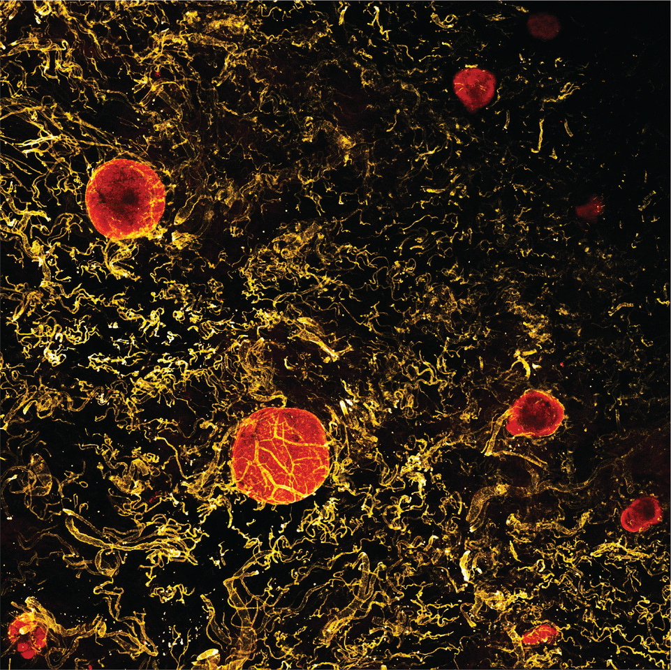
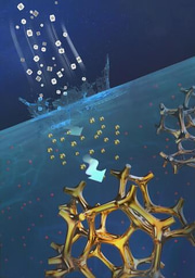
 Although we rely heavily on reductionist experiments, biology is fundamentally the study of complex systems. As such, serendipity plays a major role in biology research, and biologists must always plan to take advantage of unexpected findings wherever they arise. Our moment of serendipity came during a late-night dissection of human intestinal samples, while optimising methods for individual projects. Thomas noticed that two layers of the gut wall sometimes peeled away from one another, and attempted to reliably reproduce this to improve viability of cells after tissue digestion. After months of optimisation, we had combined this method with a staining technique used in endoscopy and, to our delight, discovered we could isolate tiny structures known as gut-associated lymphoid tissues (as visualized here by immunofluorescence microscopy - Credit to Urs M. Mörbe).
Gut-associated lymphoid tissues are one of the two major immune compartments of the gut, along with the lamina propria. The lamina propria underlies the entire surface of the fragile epithelial layer, and acts as a reactive barrier against invasive microorganisms. The gut-associated lymphoid tissues, on the other hand, are key intestinal immune priming sites and are scattered along the length of the intestines. These include the well-known appendix and Peyer’s patches, as well as numerous but small and poorly-understood clusters, known as isolated lymphoid follicles. Although these structures were first described almost two centuries ago, it has not been possible to physically separate isolated lymphoid follicles from the lamina propria in which they are partially embedded, until now. This has left a gap in our understanding of the intestinal immune system, especially in the large intestine which is rich in isolated lymphoid follicles. In particular, some recent single-cell sequencing studies found that specific cells were expanded in patients with inflammatory bowel disease. However, these studies digested and analysed multiple compartments of the gut wall together, so the expanding cells might simply reflect enlarged contaminating isolated lymphoid follicles. Our techniques now allow for high-throughput single-cell analysis of discrete human intestinal immune compartments, including uncontaminated lamina propria and the isolated lymphoid follicles.
Perhaps the most notable aspect of this story is that these new techniques do not involve highly sophisticated equipment; the key discovery was simply peeling two tissue layers apart under a dissecting microscope. It is reassuring to note that in the 21st century, when new -omics and bioinformatics technologies are published weekly, it is still possible to make meaningful discoveries with just curiosity, a supportive research environment, and a basic stereo microscope.
Follow the Topic
-
Nature Protocols

This journal publishes secondary research articles and covers new techniques and technologies, as well as established methods, used in all fields of the biological, chemical and clinical sciences.

Please sign in or register for FREE
If you are a registered user on Research Communities by Springer Nature, please sign in