Raman spectroscopy for cancer cytopathology
Published in Protocols & Methods
Cytopathology involves the morphological analysis of exfoliated cells from smears, scrapings, brushings and washings for cancer screening and investigation of suspicious lesions. For example, the Pap smear test is well established for cervical screening programmes but is known to suffer from poor sensitivity as it relies on morphological abnormalities and there may only be a few abnormal appearing cells on the slide, which can easily be missed.
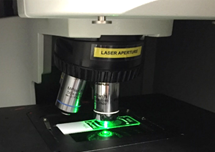
Recently, optical spectroscopic techniques have shown great promise for cancer diagnosis using tissues, cells and biofluids. In particular, Raman spectroscopy, which is an optical method based on inelastic light scattering, can provide a “biochemical fingerprint” of a sample with contributions from all the biomolecular components, including nucleic acids, proteins, lipids and carbohydrates. Raman spectra can be recorded in a rapid, label free and non-destructive manner.
In contrast to ‘spectral histopathology’, ‘spectral cytopathology’ has received less attention, perhaps due to issues with contaminants, such as the presence of blood or mucus in the sample of exfoliated cells, and the problem of finding the rare abnormal cells on unstained slides. In addition, for clinical translation, an important consideration is how Raman spectroscopy can fit into the current clinical workflow. For high precision research purposes, spectroscopic substrates, such as calcium fluoride and reflective silver coated or low e slides are commonly used to reduce the presence of spectral contributions from the substrate. However, these substrates are significantly more expensive than standard glass slides and thus not feasible for routine clinical applications such as cytopathology.
Our own recent studies have sought to address and overcome these challenges and we have shown promising results, achieving over 90% sensitivity and specificity, for identification of pre-cancer cells in cervical, oral and lung exfoliative cytology samples.
Critically, we have found that Raman spectroscopy can detect biochemical changes in morphologically normal appearing cells as a result of a field change in the whole epithelium at a biochemical level. This means that spectra can be recorded from any cells on the slide and these spectra will show biochemical features of pre-cancer.
However, a standardised approach for Raman spectral cytopathology has not yet been established. Our goal in writing this protocol was to bring together our experiences in working with a range of different cytology samples over the past few years to develop a robust standardised clinical protocol for Raman spectral cytopathology.
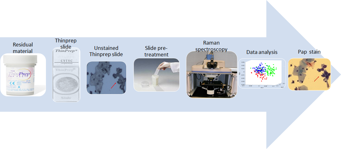
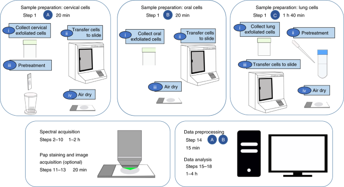
Our protocol covers sample preparation, spectral acquisition, pre-processing and data analysis. A key feature is that the protocol uses the same sample preparation procedure of liquid based cytology onto glass slides that is commonly used in cytology laboratories. This ensures compatibility with the clinical workflow. Another important feature is that the protocol includes methods of correction of the glass spectral contribution and sample pre-treatment methods to remove contaminants, such as blood and mucus, that can obscure spectral features in the exfoliated cells and lead to variability.
We hope that our protocol can be used by others for similar research on early detection of cancer using Raman spectroscopy and minimally invasive exfoliated cells.
Follow the Topic
-
Nature Protocols
This journal publishes secondary research articles and covers new techniques and technologies, as well as established methods, used in all fields of the biological, chemical and clinical sciences.


Please sign in or register for FREE
If you are a registered user on Research Communities by Springer Nature, please sign in