Seeing through the skull with 3D ultrasound matrix imaging
Published in Physics
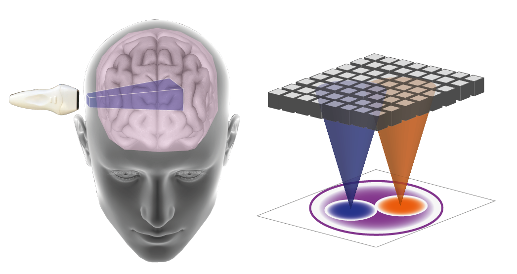

In medical echography, ultrasonic waves are used to image the human body, for applications ranging from obstetrics, vascular medicine, or hepatology to name a few. The human brain however, well-protected as it is inside the cranial vault, is very difficult to reach and remains hidden to conventional ultrasound imaging (Fig. 1).
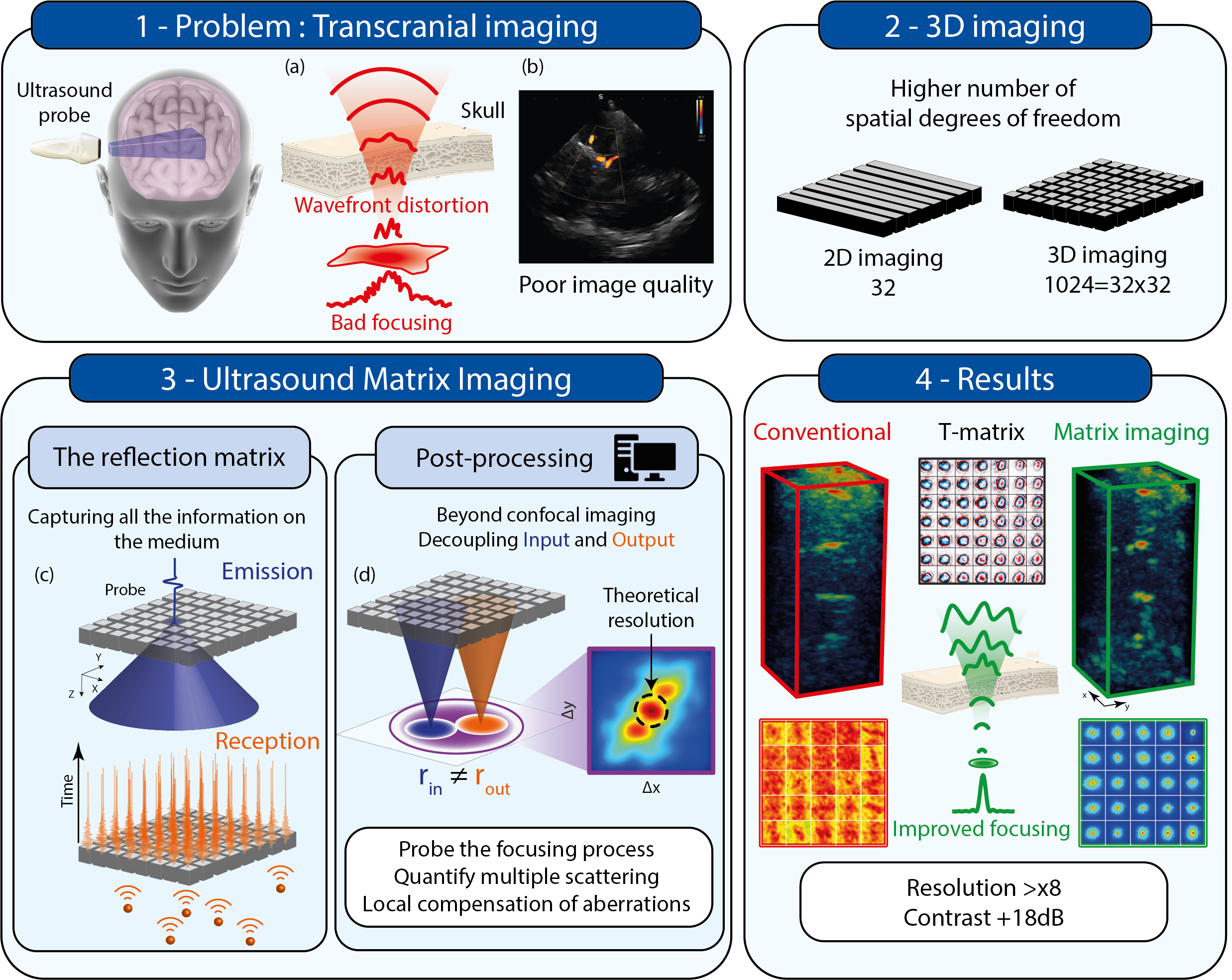
Image credit: Kjpargeter on Freepik
For any wave-based imaging technique, creating a clear image hinges on our ability to focus the wave energy at a specific point within the target medium and record the response originating from that precise location. This process is known as confocal imaging. The quality of the resulting image is heavily dependent on our ability to maintain a tight and well-controlled focus. A small focal spot with high amplitude leads to images with excellent resolution and contrast, crucial for accurate diagnosis. Unfortunately, the size of this focal spot is typically constrained by both diffraction and the irregularities in the medium through which the wave travels, especially variations in wave propagation speed. These irregularities can distort the wavefronts, resulting in a large and aberrated focal spot, ultimately leading to images with poor resolution and contrast. This explains why conventional ultrasound struggles to visualize the intricate structures of the brain and cerebral vasculature. The complex nature of the skull, combined with the exceptionally high speed of sound in bone (~3000 m/s), compared to soft tissue (~1450 m/s in the brain), introduces severe aberrations into the wavefronts, making precise focusing impossible (Fig. 1).
In this paper, we present a novel approach to ultrasound imaging, known as Ultrasound Matrix Imaging (UMI), and specifically its 3D variant (Fig. 2), which can address and rectify these aberrations, enabling us to see through the skull.
The crux of our method involves acquiring something we term the "Reflection Matrix," which serves as a way to capture extensive information about the medium we wish to image. To achieve this, we emit short pulses from every transducer element that makes up the ultrasound probe and record the responses reflected back by the medium on all these transducer elements. This approach yields a substantial amount of data. Instead of focusing on a single point and listening to that point's response, we employ unfocused waves that illuminate the entire medium. We then listen to the responses from all points in the medium simultaneously (Fig. 3). However, this data can be quite complex and challenging to analyze, given that it comes from every point within the medium. Thus, the trick is to sort and organize this information to extract the quantitative data of interest.
Our solution involves applying specific combinations of delays and summations to all the elements within the Reflection Matrix to transform it into a "Focused Reflection Matrix." While this name might suggest a return to conventional confocal imaging, it's much more than that. Since we've gathered all available information about the medium, we can not only virtually focus on any given point but also recover the response from this point as well as from any other point in the medium. In essence, we can virtually focus on distinct points during both emission and reception, allowing us to assess focus quality by extracting the Reflection Point Spread Function (RPSF, see Fig. 3). For a given emission focal spot, this RPSF illustrates how much energy is reflected by nearby points. In an ideal setting with no aberrations, the RPSF size is determined by diffraction: we focus the wave over an area roughly the size of the wavelength in both emission and reception, containing all reflected energy within the black dotted circle in Fig. 3. However, in the presence of aberrations, the reflected energy scatters over a larger area. The RPSF thus serves as an indicator of image quality and can be used to correct aberrations.
To validate our approach, we conducted experiments using a head phantom (Fig. 4), which mimics the challenges of brain imaging through the skull bone. Our results demonstrate that Ultrasound Matrix Imaging effectively corrects skull-induced aberrations, resulting in a clear and tightly focused diffraction-limited image. This correction significantly improves the resolution and contrast compared to conventional confocal imaging methods.
Flavien Bureau, Justine Robin & Alexandre Aubry
Follow the Topic
-
Nature Communications
An open access, multidisciplinary journal dedicated to publishing high-quality research in all areas of the biological, health, physical, chemical and Earth sciences.

Related Collections
With Collections, you can get published faster and increase your visibility.
Women's Health
Publishing Model: Hybrid
Deadline: Ongoing
Tumor Microenvironment Crosstalk and Therapeutic Implications
Publishing Model: Hybrid
Deadline: Nov 02, 2026

Please sign in or register for FREE
If you are a registered user on Research Communities by Springer Nature, please sign in