Using physics to test corneal cell quality
Published in Bioengineering & Biotechnology
Our eyes sometimes need significant repair, like cornea transplantations. Using one equation in colloid physics, we assess the collective cell order of corneal endothelial cells on a dish or in the patients’ eyes after transplantation, which helps clinicians intervene before severe symptoms appear.
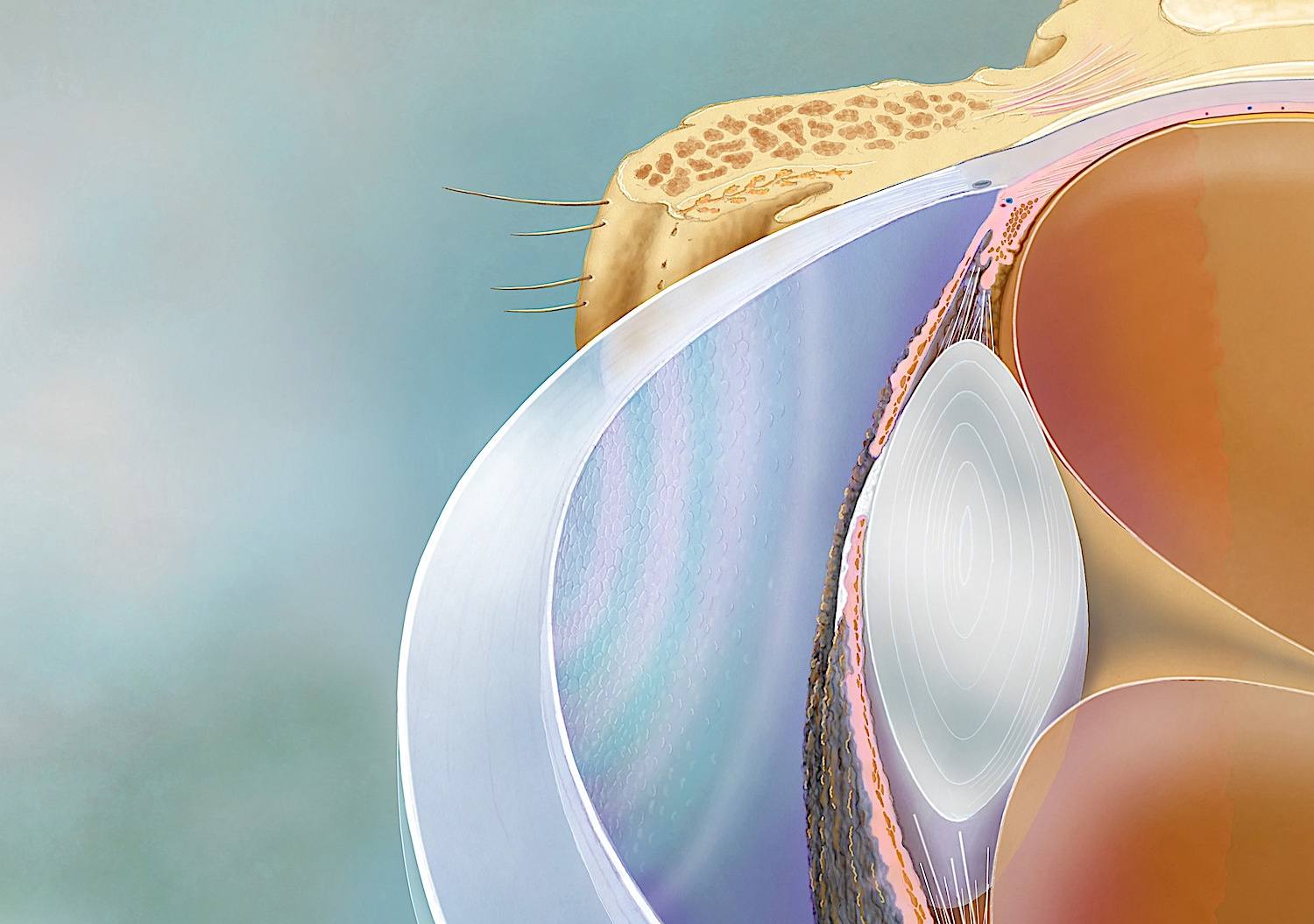
Our eyes — the windows to the soul — need constant care, and as we age, they sometimes also need significant repair. Now thanks to our team in Kyoto, transplanting the panes of these windows, the corneas, may become even safer.
My colleagues and I, led by Kyoto University physicists and Kyoto Prefectural University of Medicine (KPUM) ophthalmologists, have developed a quantitative biomarker that makes it possible to assess the quality of corneal cells — and even predict their long-term efficacy — through simple observation.
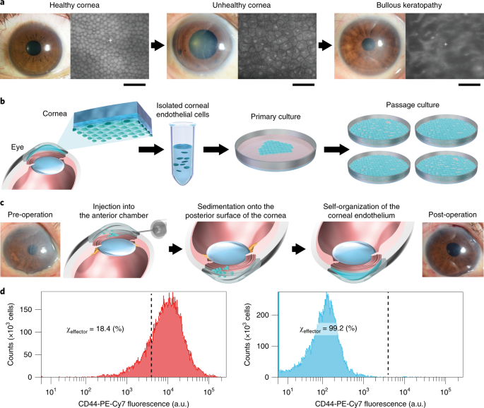
Cornea transplantations generally become necessary when endothelial cells decrease in number, resulting in haziness. But endothelia don’t multiply well in the body, which is why there has been a need to rely on the transplantation of donor corneas for treatment.
Fortunately, in 2009, my colleagues at KPUM succeeded in developing a method to culture the cells in a dish. These new cells could then be then transplanted into the eyes of patients and restore their corneas to health.
This method has shown significant promise in clinical trials, but two major obstacles to wider application have remained: quality control of cells before injection and confirmation of long-term functionality.
Typically, cell quality is assessed through protein expression patterns via flow cytometry. However, a single test requires almost 100,000 cells and relies heavily on the observations and experience of senior professionals.
Cells in a tissue constantly interact with each other to maintain homeostasis, which was the hint we needed to employ colloid physics — used for measuring interactions of micro- and nanoparticles — in a new method to assess the cornea cells. Calculating the interactions between all cells in the cornea allowed us to find the spring constant, correlating with collective cell order.
Assessment is relatively simple. We only need to extract the rims of the cells, either from a microscopic image in a culture dish or from ophthalmological inspection images of the patients’ eyes. Both the quality of the cells and their long-term efficacy can be determined with just one equation.
The procedure has potential applications in preemptive medicine, enabling clinicians and doctors to intervene before more severe symptoms appear. We foresee that our quantitative biomarker, and the concept behind it, will be applied to other epithelial cell cultures and tissues in the future.
The paper “A physical biomarker of the quality of cultured corneal endothelial cells and of the long-term prognosis of corneal restoration in patients” appeared on 22 July 2019 in Nature Biomedical Engineering, with doi: 10.1038/s41551-019-0429-9
Follow the Topic
-
Nature Biomedical Engineering
This journal aspires to become the most prominent publishing venue in biomedical engineering by bringing together the most important advances in the discipline, enhancing their visibility, and providing overviews of the state of the art in each field.

Related Collections
With Collections, you can get published faster and increase your visibility.
Implantable wireless communication technologies
Publishing Model: Hybrid
Deadline: Nov 28, 2026
Medical Ultrasound: Emerging Techniques and Applications
Publishing Model: Hybrid
Deadline: Jan 29, 2027

Please sign in or register for FREE
If you are a registered user on Research Communities by Springer Nature, please sign in