When Pemphigus Foliaceus Breaks the Rules: Two Rare Cases of Pustular Disease That Challenged the Diagnosis
Published in Biomedical Research
Explore the Research
Atypical pemphigus foliaceus with pustular lesions: a case series
When appearances deceive in autoimmune blistering disease
One of the greatest challenges in dermatology is recognizing when a familiar disease presents in an unfamiliar way. Pemphigus foliaceus (PF) is classically characterized by superficial erosions, crusts, and scaling resulting from autoantibodies directed against desmoglein 1. Clinicians generally do not expect prominent pustules to dominate the presentation.
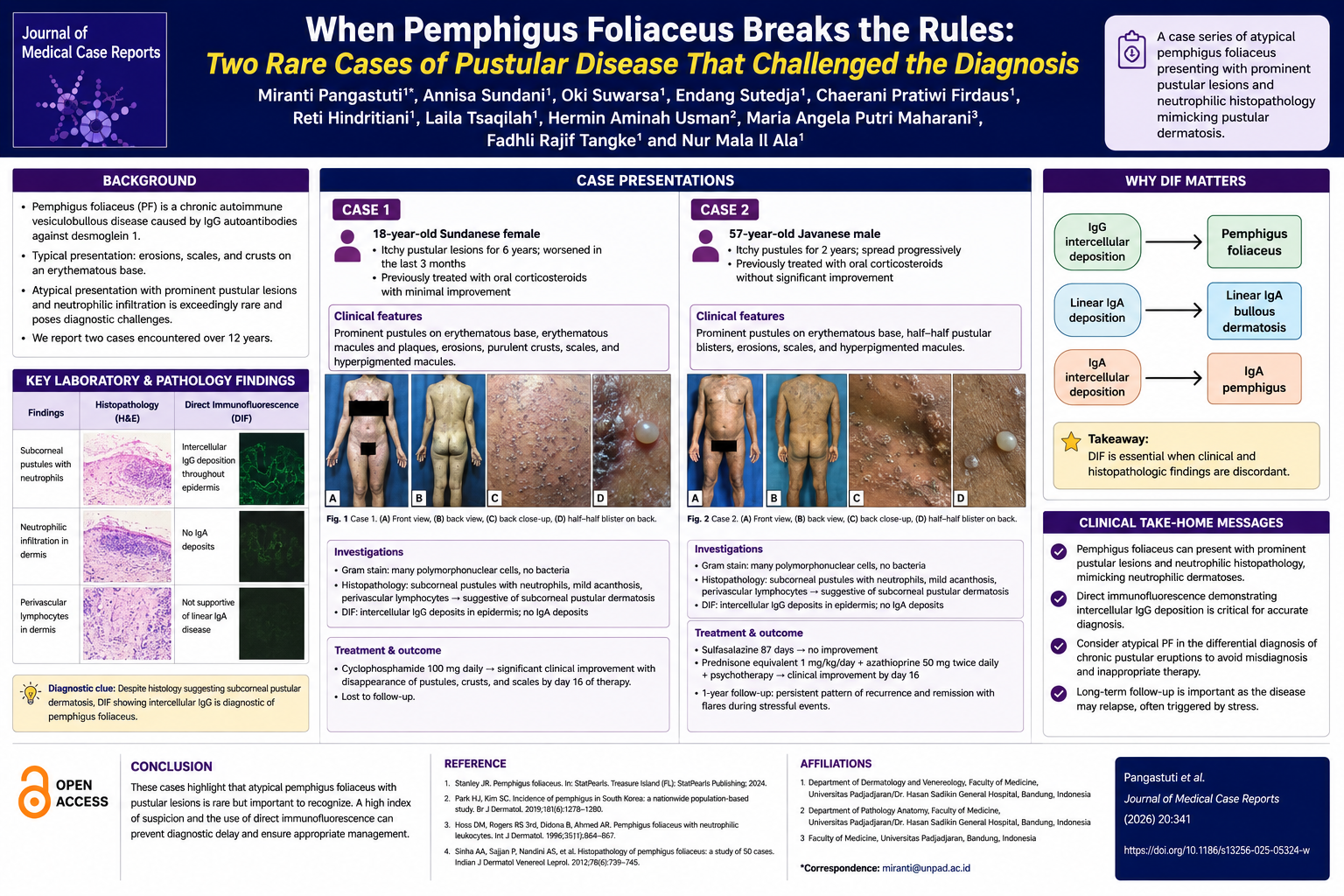
In this Journal of Medical Case Reports case series, Pangastuti and colleagues describe two patients whose clinical appearance initially suggested an entirely different disease process. Both presented with extensive pustular eruptions accompanied by neutrophilic infiltration on histopathology—features far more consistent with subcorneal pustular dermatosis than classic PF.
The first patient was an 18-year-old woman with a 6-year history of progressive pruritic pustular lesions that had failed multiple courses of corticosteroids. The second was a 57-year-old man with a 2-year history of recurrent pustular eruptions that likewise proved resistant to conventional therapy. In both patients, Gram stains demonstrated abundant neutrophils without bacteria, and histopathology revealed subcorneal pustules dominated by neutrophilic inflammation, reinforcing the initial impression of a neutrophilic dermatosis.
The diagnostic turning point came with direct immunofluorescence (DIF). Rather than demonstrating IgA deposition typical of IgA pemphigus or subcorneal pustular dermatosis, both specimens showed intercellular IgG deposition throughout the epidermis—a hallmark of pemphigus foliaceus. This finding established the correct diagnosis despite the highly atypical morphology.
These cases illustrate an important diagnostic principle: histopathology alone may not be sufficient when autoimmune blistering disorders present outside their classic phenotype. The authors emphasize that DIF should remain an essential component of the evaluation whenever chronic pustular eruptions remain unexplained or fail to respond as expected.
Management also reflected the complexity of these cases. One patient experienced rapid improvement with cyclophosphamide after prolonged unsuccessful corticosteroid treatment. The second improved after treatment with systemic corticosteroids and azathioprine, together with psychotherapy, highlighting both the chronic relapsing nature of the disease and the importance of addressing psychological burden alongside immunosuppressive therapy.
Although only two cases, this series expands the recognized clinical spectrum of pemphigus foliaceus. Rare presentations such as these remind clinicians that autoimmune blistering diseases can masquerade as neutrophilic dermatoses, making comprehensive clinicopathologic correlation indispensable.
For dermatologists, dermatopathologists, and clinicians evaluating chronic pustular disorders, the report reinforces a simple but valuable lesson: when the clinical picture and histology do not fully agree, direct immunofluorescence may provide the decisive answer.
Clinical Take-Home Message
Atypical pemphigus foliaceus may present predominantly with pustular lesions and neutrophilic histopathology, closely mimicking subcorneal pustular dermatosis. When clinical and histologic findings are discordant, direct immunofluorescence demonstrating intercellular IgG deposition is critical for establishing the diagnosis and guiding appropriate immunosuppressive therapy.
Multiple-Choice Question
A patient presents with chronic, widespread pustular skin lesions and histopathology demonstrates subcorneal pustules with prominent neutrophilic infiltration, suggesting subcorneal pustular dermatosis. Which diagnostic finding is most important for establishing the diagnosis of atypical pemphigus foliaceus?
A. Positive bacterial culture from pustular fluid
B. Linear IgA deposition along the basement membrane on direct immunofluorescence
C. Intercellular IgG deposition on keratinocyte surfaces demonstrated by direct immunofluorescence
D. Peripheral blood eosinophilia with elevated serum IgE
Correct Answer: C. Intercellular IgG deposition on keratinocyte surfaces demonstrated by direct immunofluorescence
Explanation
Why C is correct:
The hallmark diagnostic feature of pemphigus foliaceus is intercellular IgG deposition within the epidermis on direct immunofluorescence (DIF). In the reported cases, both patients had atypical pustular lesions and neutrophilic histopathology that initially mimicked subcorneal pustular dermatosis. However, DIF demonstrated the characteristic intercellular IgG staining pattern, confirming the diagnosis of atypical pemphigus foliaceus despite its unusual clinical presentation.
Why the other answers are incorrect:
- A. Positive bacterial culture from pustular fluid – Incorrect. Both patients had numerous neutrophils on Gram stain but no bacteria, arguing against an infectious pustular process.
- B. Linear IgA deposition along the basement membrane – Incorrect. Linear IgA deposition is characteristic of linear IgA bullous dermatosis, not pemphigus foliaceus. The reported cases specifically lacked IgA deposition on DIF.
- D. Peripheral blood eosinophilia with elevated serum IgE – Incorrect. Although eosinophils may be seen in some inflammatory skin diseases, peripheral eosinophilia and elevated IgE are not diagnostic criteria for pemphigus foliaceus and were not the key findings in these cases.
Educational Pearl:
When chronic pustular eruptions have histopathologic features suggestive of neutrophilic dermatoses but the clinical picture remains atypical, direct immunofluorescence is essential. Intercellular epidermal IgG deposition can distinguish atypical pemphigus foliaceus from other pustular dermatoses and prevent delayed diagnosis and inappropriate treatment.
Journal of Medical Case Reports is the world's first international, PubMed-listed medical journal devoted to publishing case reports from all medical disciplines and will consider any original case report that expands the field of general medical knowledge, and original research relating to case reports. The journal is open access and strongly endorses the CARE guidelines for case reports, requiring authors to submit populated CARE checklists with submissions to improve transparency in reporting.

Richard Alan Rison is the interim Editor-in-Chief of Journal of Medical Case reports. He is also an associate neurology editor (editorial board) for BMC Neurology, and the former lead editor for case reports of BMC Research Notes (currently on the editorial board). His scholarly work focuses on medical case reporting, reporting standards, and editorial methodology. Dr. Rison participated in the development and dissemination of the CARE guidelines for clinical case reporting and has authored numerous publications addressing both neurological disorders and the role of case reports in advancing medical knowledge. Dr. Rison practices general neurology and served as the founding medical director of the PIH Health Hospital-Whittier Stroke Program and the PIH Health Hospital-Whittier Non-Invasive Vascular Laboratory, is a clinical assistant professor of neurology at the University of Southern California Keck School of Medicine and Los Angeles County Medical Center, and is a Fellow of the American Academy of Neurology, the American Neurological Association, and the American Association of Neuromuscular and Electrodiagnostic Medicine. Dr Rison is board-certified by the American Board of Psychiatry and Neurology in neurology and vascular neurology, and neurocritical care and neuroimaging by the United Council of Neurologic Subspecialties. He is also board-certified by the American Board of Electrodiagnostic Medicine in electrodiagnostic medicine. Dr. Rison is a former president of the Los Angeles Neurological Society.
Follow the Topic
-
Journal of Medical Case Reports
This journal will consider any original case report that expands the field of general medical knowledge, and original research relating to case reports.




Please sign in or register for FREE
If you are a registered user on Research Communities by Springer Nature, please sign in