A New Way to Characterize Cellular Heterogeneity in Tumours
Published in Bioengineering & Biotechnology

Tumours consist of heterogeneous populations of cancer cells that have distinct genetic and phenotypic profiles. Cellular heterogeneity within a tumour, namely intratumoural heterogeneity, has become a great barrier to effective cancer therapy. Assessing the extent of intratumoural metabolic heterogeneity would greatly contribute to our understanding of tumour growth, invasion, and drug resistance. It will also help design effective and personalized treatment strategies by predicting sensitivity or resistance.
The oxygen consumption rate (OCR) of a cell is directly related to its metabolism. The distribution of single-cell OCRs within a tumour is an important gauge of intratumoural metabolic heterogeneity. Thus far, single-cell OCRs have been measured by electrical and fluorescent methods, which require microfabricated oxygen sensors to monitor the oxygen content change. However, these two methods are difficult to scale up for measuring a large number of cells. Moreover, the embedded oxygen sensors may adversely affect the normal metabolism of the cells, rendering the OCR measurement inaccurate.
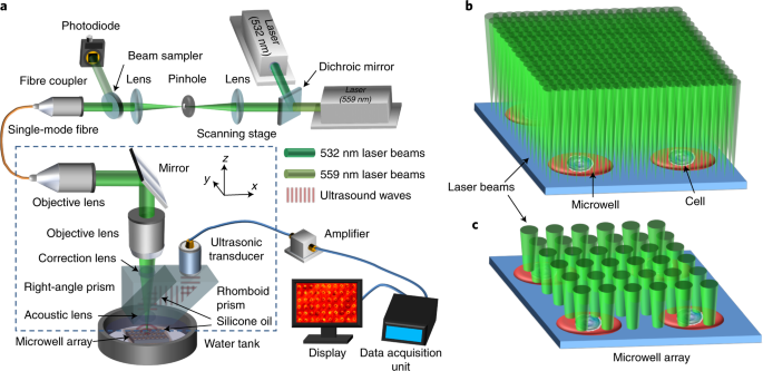
To address this issue, we turned our attention to haemoglobin, the natural oxygen carrier and supplier for cells in animal tissues. Significant color changes occur when haemoglobin adsorbs or desorbs oxygen because its optical absorption spectrum varies when it becomes oxygenated and deoxygenated. This unique property makes haemoglobin an ideal biocompatible optical-absorption-based oxygen sensor. Combining with photoacoustic microscopy, the most sensitive imaging technology for detecting change in optical absorption, we were able to monitor tiny variations in oxygen saturation of haemoglobin, from which the OCR of each cell can be derived. We termed this technique as single-cell metabolic photoacoustic microscopy (SCM-PAM). Without the need of microfabricated oxygen sensors, SCM-PAM could be easily scaled up for measuring a large population of single cells.
To ensure accurate single-cell OCR measurement, each cell and a small amount of haemoglobin (solution) must be sealed inside a small oxygen-diffusion-limited microwell. In the beginning, this requirement appeared to be extremely challenging to meet. To ensure a high yield of single cell entrapment, each microwell must be made just slightly larger than the average size of cells. However, such a tight fit leaves little room for adding haemoglobin. To address this issue, we replaced the glass microwell substrate with an anodisc filter. With numerous through nanopores, the anodisc filter can hold quite a bit of water solution like a sponge. What is more, because the nanopores are isolated from each other, individual oxygen-diffusion-limited microwells (each consisting of a separated cell chamber and oxygen supply/sensing chamber) will be automatically formed once the top and bottom surfaces of the microwell array are sealed, say, with a thin layer of oil.
So far, we have performed single-cell OCR measurements on normal and cancer cells from both cultured cell lines and breast cancer patients under different physiological conditions. Our preliminary statistical analysis clearly indicates the escalated metabolic heterogeneity of cancer cells compared with normal ones. With its unique capability for label-free high-throughput single-cell OCR measurement and the potential of providing multidimensional information about tumours, we believe SCM-PAM will become a useful tool for both fundamental cancer research and clinical personalized cancer therapy.
Written by Jun Zou (junzou@ece.tamu.edu) and Lihong V. Wang (lihong@caltech.edu).
Our paper: Pengfei Hai, Toru Imai, Song Xu, Ruiying Zhang, Rebecca L. Aft, Jun Zou, and Lihong V. Wang, “High-throughput, label-free, single-cell photoacoustic microscopy of intratumoral metabolic heterogeneity,” Nature Biomedical Engineering (2019) DOI: https://doi.org/10.1038/s41551-019-0376-5.
Follow the Topic
-
Nature Biomedical Engineering

This journal aspires to become the most prominent publishing venue in biomedical engineering by bringing together the most important advances in the discipline, enhancing their visibility, and providing overviews of the state of the art in each field.
Related Collections
With Collections, you can get published faster and increase your visibility.
Implantable wireless communication technologies
Publishing Model: Hybrid
Deadline: Nov 28, 2026
Biosensing
Publishing Model: Hybrid
Deadline: Jun 30, 2026

Please sign in or register for FREE
If you are a registered user on Research Communities by Springer Nature, please sign in