A Quantitative Analysis of Ligand Binding at the Protein-Lipid Bilayer Interface
Published in Chemistry, Physics, and Cell & Molecular Biology
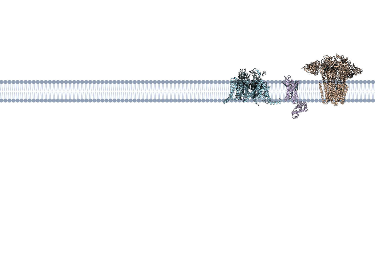
When you take a drug, chances are it's designed to interact with a specific protein in your body. Many of these proteins are embedded inside membranes called lipid bilayers. A lipid bilayer is made up of two layers of molecules called phospholipids, which have two distinct parts: a hydrophilic (water-attracting) head and a hydrophobic (water-repelling) tail. Proteins embedded in the lipid bilayer play important roles in many biological processes, such as sending signals between cells or enabling sensory functions like touch. Because they are involved in so many functions, membrane proteins are prime targets for drugs treating conditions like heart disease, pain, and many others. In fact, the majority of drugs bind to membrane proteins1!
Many drug discovery efforts focus on targeting the parts of membrane proteins that are exposed to water, rather than the parts that touch the lipid bilayer2. However, these water-exposed sites may be too similar across different proteins, leading to unintended side effects when drugs bind to multiple targets3. We were interested in understanding how the properties of molecules that bind to lipid-exposed sites differ from those that interact with water-exposed regions. To do this, we collected a dataset of molecules that bind at the protein-lipid bilayer interface, consisting of 3D structures obtained through imaging techniques like X-ray crystallography and cryo-electron microscopy. Our dataset is called the Lipid-Interacting LigAnd Complexes Database (LILAC-DB) and contains 413 structures of molecules that bind to proteins at the protein-lipid bilayer interface. 11% of molecules in the dataset are FDA approved drugs and 9% of the molecules are in clinical trials.
The dataset is publicly available to download at https://zenodo.org/records/14835079.
 bound to Cinacalcet (PDB ID: 7M3F). CaSR is shown in purple with a yellow cylinder indicating the region surrounded by the hydrophobic regions of lipids and blue cylinders indicating regions surrounded by water. Cinacalcet is shown in pink.")
One example that highlights the importance of lipid-exposed drug binding is the calcium-sensing receptor (CaSR)4, shown in purple in Animation 1, with blue cylinders marking where water is located and yellow cylinders representing where the hydrophobic part of lipid molecules are located. CaSR is a membrane protein that helps regulate calcium levels in the body. CaSR detects calcium levels in the bloodstream, triggering signals that control the release of parathyroid hormone. In some conditions, such as hyperparathyroidism, CaSR function becomes dysregulated, causing excessive calcium release. To treat this, scientists have developed drugs like cinacalcet (shown in pink in Animation 1). Cinacalcet binds to CaSR, making it more responsive to calcium signals5. Unlike many drugs that bind to water-exposed regions of proteins, cinacalcet binds at a site touching the lipid bilayer, making it an excellent example of how membrane-facing binding sites can be successfully targeted in drug design.
Key Findings from the Study
Analyzing all of the systems in LILAC-DB, we found that molecules binding to lipid-exposed sites tend to be larger, more fat-soluble, and contain more halogen atoms (such as the fluorine atoms shown in green in Animation 1) than drugs that bind to water-exposed protein regions. These properties help the molecules interact effectively with the fatty environment of the membrane. Understanding these trends will help scientists prioritize what types of molecules to test when they are designing new molecules that bind to lipid-facing sites.
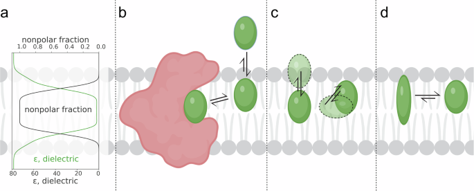
We also learned that the position of a molecule within the membrane affects its chemical characteristics. Molecules deeper in the membrane tend to have different atomic properties than those near the surface; for example, parts of molecules embedded deep in the hydrophobic part of the lipid bilayer tend to have a lower magnitude of partial charge than those interacting with water. This finding suggests that drug properties need to be tuned to match their intended location within the membrane, which will help scientists design new drugs.
Proteins are made up of building blocks called amino acids, each with unique chemical properties that determine how a protein folds and functions. We learned that lipid-exposed drug-binding sites tend to have a different mix of amino acids than those exposed to water. Understanding these patterns will help scientists predict where potential drug-binding sites might be located on membrane proteins, aiding in the discovery of new drug targets.
Understanding how molecules interact with membrane proteins is important for the discovery of new drugs. Our study provides insight into the unique opportunities and challenges posed by lipid-facing binding sites. Unlike water-exposed regions, which are often targeted in traditional drug discovery efforts, lipid-exposed regions present distinct chemical environments that require drugs with specific properties. Because the “rules” of drug discovery may be different at lipid-exposed sites, understanding these molecular interactions can help guide the design of drugs that effectively target membrane proteins. The LILAC-DB is a useful resource for studying membrane-associated drug binding and will contribute to new approaches in designing drugs which bind to membrane proteins.
References
- Santos, R. et al. A comprehensive map of molecular drug targets. Nat. Rev. Drug Discov. 16, 19–34 (2017).
- Christopoulos, A. Allosteric binding sites on cell-surface receptors: novel targets for drug discovery. Nat. Rev. Drug Discov. 1, 198–210 (2002).
- Owen, R. M. et al. Design and Identification of a Novel, Functionally Subtype Selective GABAA Positive Allosteric Modulator (PF-06372865). J. Med. Chem. 62, 5773–5796 (2019).
- Gao, Y. et al. Asymmetric activation of the calcium-sensing receptor homodimer. Nature 595, 455–459 (2021).
- The calcium-sensing receptor in physiology and in calcitropic and noncalcitropic diseases | Nature Reviews Endocrinology. https://www.nature.com/articles/s41574-018-0115-0#Sec20.
Follow the Topic
-
Communications Chemistry

An open access journal from Nature Portfolio publishing high-quality research, reviews and commentary in all areas of the chemical sciences.
Related Collections
With Collections, you can get published faster and increase your visibility.
Experimental and computational methodology in structural biology
Publishing Model: Open Access
Deadline: Apr 30, 2026
Advances in Asymmetric Catalysis for Organic Chemistry
Publishing Model: Open Access
Deadline: Mar 31, 2026

Please sign in or register for FREE
If you are a registered user on Research Communities by Springer Nature, please sign in