Advancing systemic mastocytosis research through a novel pre-clinical model
Published in Biomedical Research
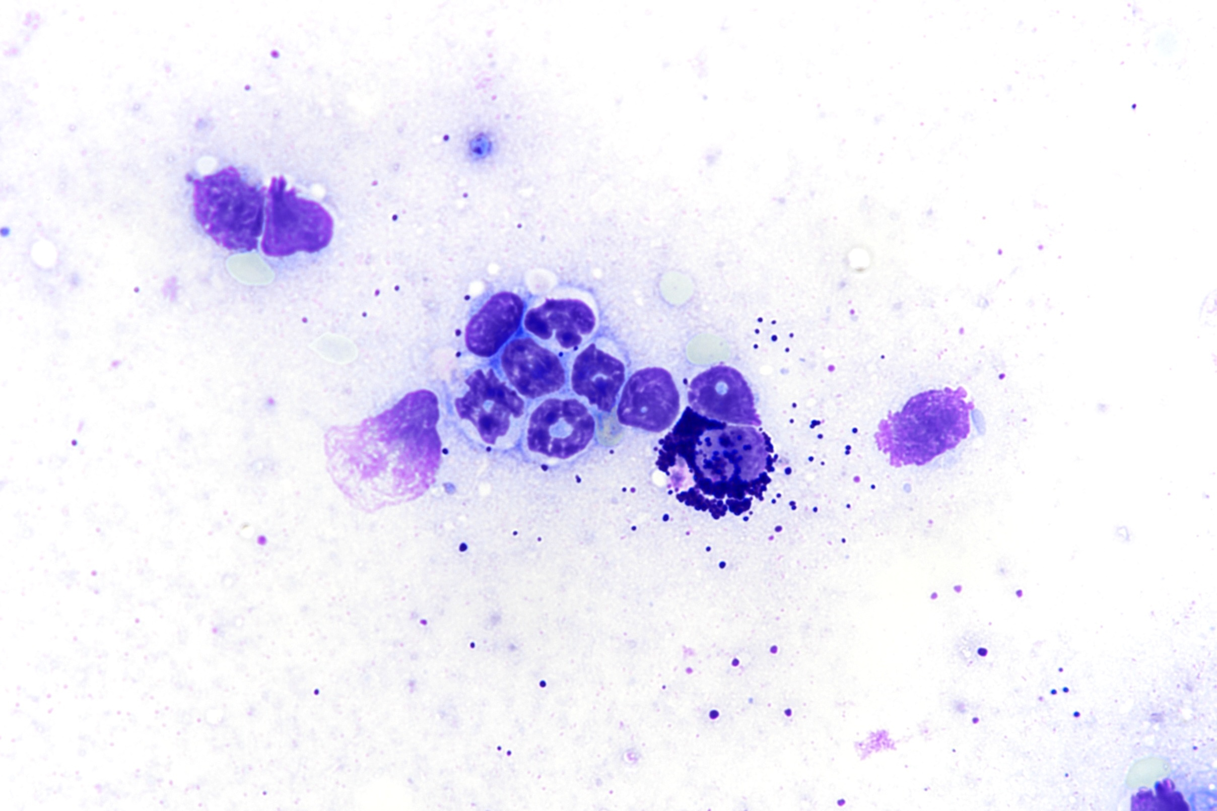

Mastocytosis is a rare disease arising as a consequence of DNA mutations in mast cells (MC), causing their uncontrolled proliferation and accumulation. Approximately 1-2 people in 100’000 are detected every year, but its diagnosis is often difficult and lengthy due to the very heterogeneous presentation, ranging from only cutaneous involvement in the most benign forms to the advanced cases of systemic mastocytosis or mast cell leukemia, with very poor prognosis. This array of clinical symptoms isn’t the results of a diverse genetic cause; on the contrary, the molecular alteration driving mastocytosis is in the majority of cases a single mutation located in the gene coding for the receptor tyrosine kinase Kit. As a consequence, the receptor is active even in absence of its ligand. Because mast cells rely on Kit signaling for their survival and proliferation, the presence of the activating mutation (D816V in exon 17) causes them to proliferate resulting in their accumulation not just in those organs where they are normally present (such as the skin, the stomach and intestine, or the lungs), but also in other tissues, such as the bone marrow, spleen, or liver. On the long term, infiltration and accumulation of mast cells can lead to organ damage and malfunction, as a result of the release of mediators and bioactive molecules stored in their cytoplasmatic granules. While for many years treatment of mastocytosis only relied on symptoms handling and prevention of life-threatening anaphylaxis, the discovery of the Kit D816V mutation has opened the way for the development of specific tyrosine kinase inhibitors. The approval of midostaurin, a multi-kinase inhibitor, and particularly of avapritinib, a Kit D816V-specific inhibitor, represented a major step forward in the treatment of mastocytosis, with deep and durable responses over long-duration treatment.
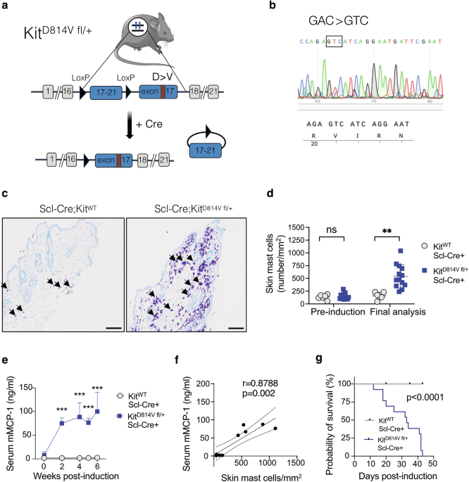
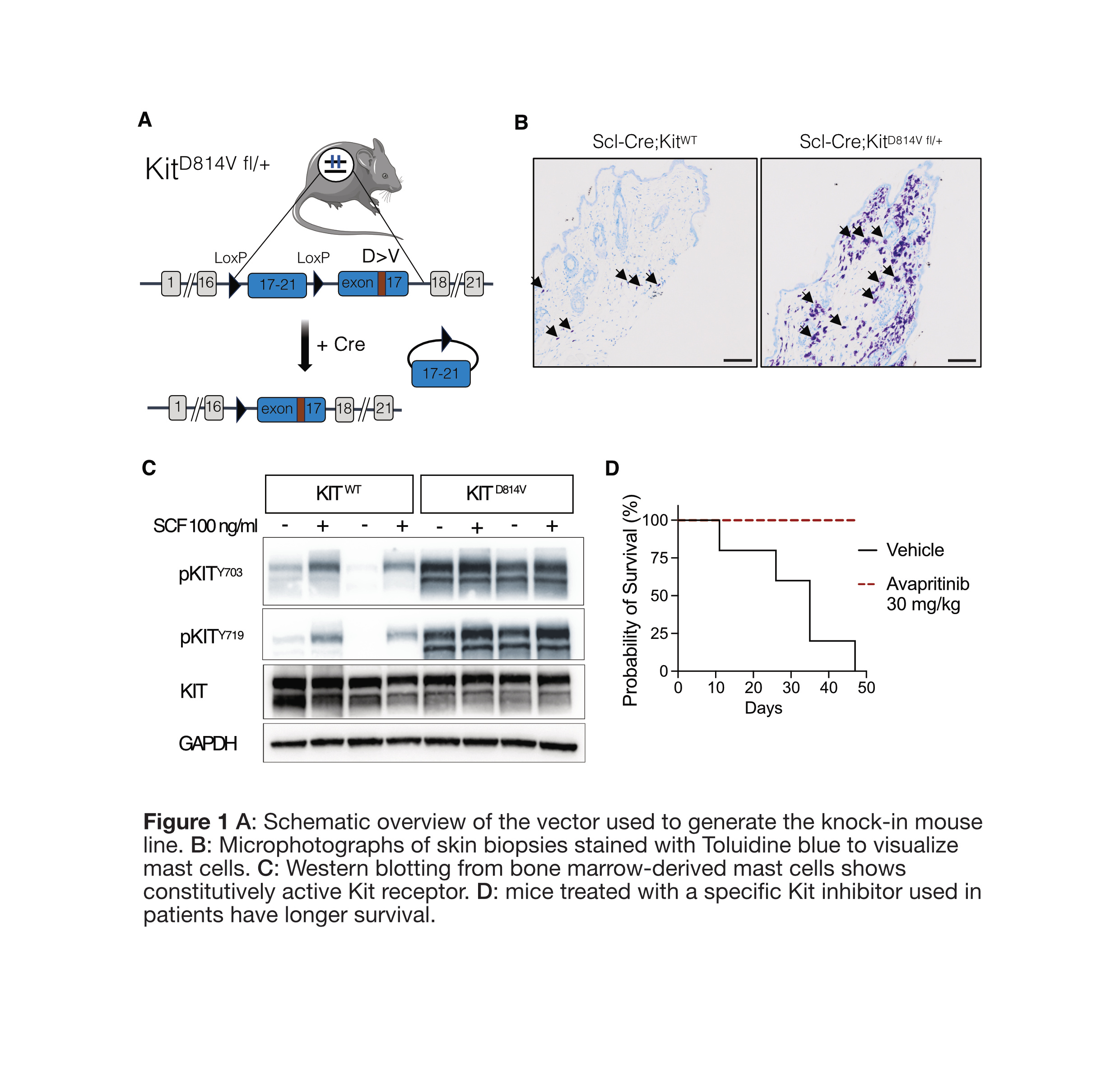
Many questions, however, remain open: how can a single mutation result in so many different manifestations in patients? Which pathways are altered in neoplastic mast cells as a result of Kit D816V, and which are responsible for which symptom(s)? What changes at the cellular level are induced in the different tissues as a result of mast cells accumulation? To be able to solve these (and many others) puzzles, good pre-clinical models are needed, that would ideally recapitulate the most important features of the disease in patients. While numerous mouse models exist for other malignancies, they have been scarce so far in the field of mastocytosis. Existing models were mostly based on cell lines-derived xenografts into immunodeficient mice, which obviously cannot recapitulate the origin of the disease and the cross-talk between the different immune cells known to play an important role in cancer. Therefore, we employed the CRISPR-Cas9 technology to generate a knock-in mouse line expressing the Kit D814V mutation (the murine equivalent of the D816V mutation found in most patients), inducing its expression in the hematopoietic compartment through recombination mediated by Scl-Cre (expressed in hematopoietic stem and progenitor cells, Figure 1A). We observed the mice over several weeks by taking periodically small blood samples and skin biopsies, which we analyzed for changes in blood values parameters and number of mast cells, respectively. We found that the mice had profound alterations in both: the number of mast cells was significantly increased in the skin of mice expressing Kit D814V (Figure 1B), and they had extremely high values of white and red blood cells (WBC and RBC). After euthanasia, we extended our analysis on several organs (bone marrow, spleen, liver, lungs), where we confirmed the presence of mast cells and their progenitors (derived from hematopoietic stem cells) in a higher number compared to mice carrying the wild-type version of Kit. The elevated blood counts were the result of extramedullary hematopoiesis, which happens when blood stem cells exit the bone marrow and migrate to the spleen and liver, where they proliferate and produce more mature cells entering the blood stream. In line with the systemic expansion of mast cells, serum levels of the respective MC protease-1 were elevated; many other cytokines were also increased in Kit D814V mice, of which some relevant pro-inflammatory molecules (TF-alpha, IL-1beta, IL-17), several chemokines and growth factors. Overall, from the data obtained we could conclude that this novel mouse line presents a phenotype of systemic mastocytosis with associated hematological neoplasm, one of the advanced forms of the disease.
An important aspect of the analysis and validation of this line as a preclinical model for mastocytosis was to demonstrate that 1) the D814V mutation results in ligand-independent activation of Kit; 2) the mastocytosis phenotype can be reversed by treatment with specific Kit inhibitors such as those administered to patients. For the first point we derived mast cells ex vivo by cultivating bone marrow cells in presence of IL-3, the cytokine directing the maturation and proliferation of MC. When their purity was >95%, cells were analyzed for phosphorylated Kit (indicating its active state) with or without activation with stem cell factor (SCF), the specific Kit ligand. Indeed, western blot analysis showed that unlike Kit WT cells, which had little phosphorylated receptor without stimulation and robustly responded to activation by SCF, cells expressing Kit D814V presented high levels of active Kit at basal level, indicating its active state independently of SCF binding (Figure 1C).
For the second point, we randomized Kit D814V mice to receive vehicle or the specific inhibitor avapritinib, which is approved for clinical use in advanced mastocytosis patients. At the end of the 2 weeks treatment, mice were euthanized and found to have lower skin mast cells, reduced serum MC protease levels, and normalized spleen size. A group of mice was treated for a longer period of time to show that avapritinib treatment increased survival, compared to vehicle-treated mice which died or had to be euthanized during the course of the treatment (Figure 1D).
The generation and initial characterization of this novel mouse line represents a major step forward in the field of mastocytosis, with which we hope to answer some of the open questions in the field and, eventually, improve the treatment of patients.

Follow the Topic
-
Leukemia
This journal publishes high quality, peer reviewed research that covers all aspects of the research and treatment of leukemia and allied diseases. Topics of interest include oncogenes, growth factors, stem cells, leukemia genomics, cell cycle, signal transduction and molecular targets for therapy.