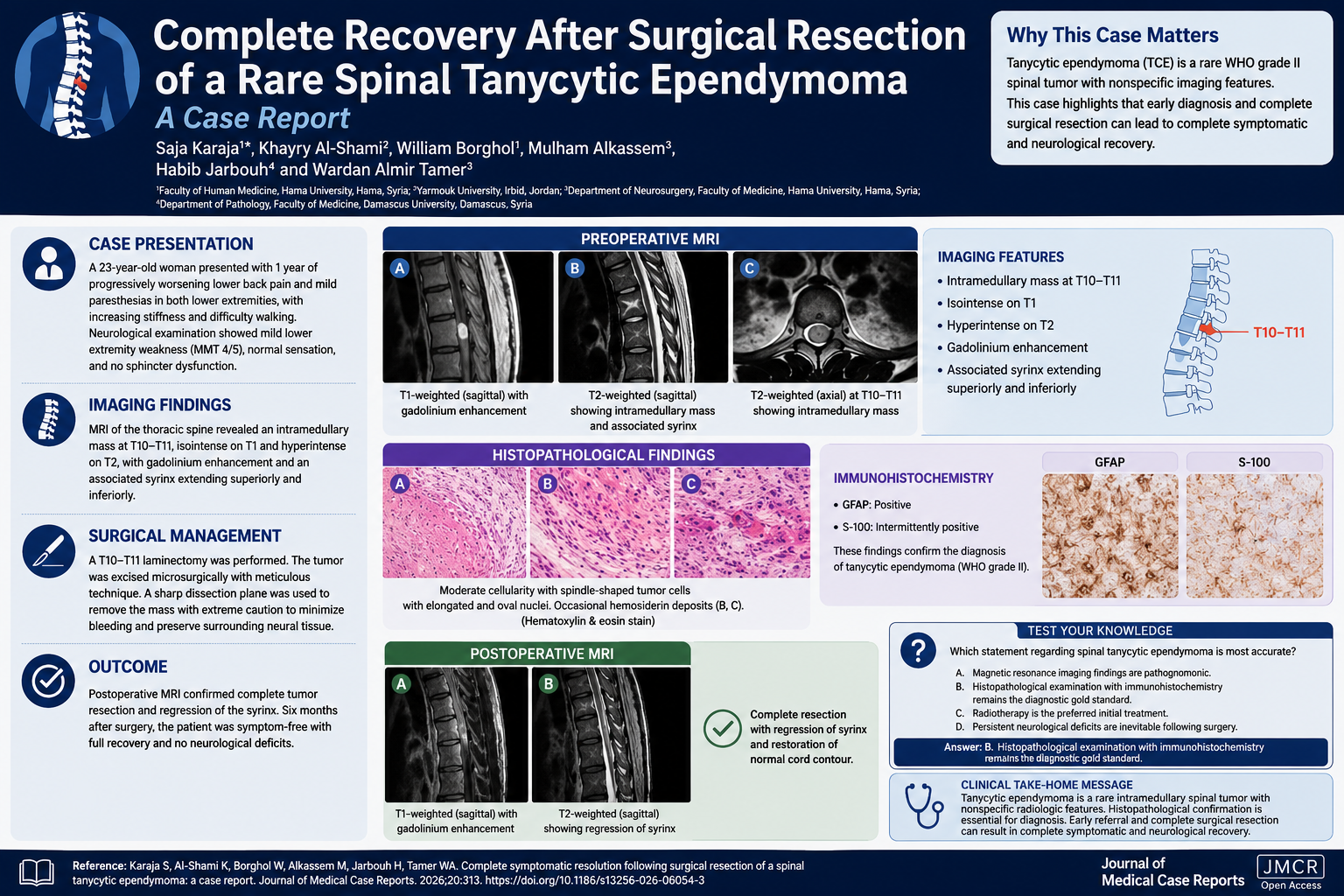
Complete Recovery After Surgical Resection of a Rare Spinal Tanycytic Ependymoma
Published in Neuroscience and Biomedical Research
Explore the Research
Complete symptomatic resolution following surgical resection of a spinal tanycytic ependymoma: a case report
Tanycytic ependymoma is an uncommon World Health Organization grade II ependymoma arising from tanycytes and occurring predominantly within the spinal cord. Because its radiologic appearance overlaps with other intramedullary tumors, establishing the diagnosis before surgery is often challenging.
Karaja and colleagues report the case of a 23-year-old Syrian woman who experienced progressive lower back pain and bilateral lower-extremity paresthesias for one year. Neurological examination demonstrated mild lower extremity weakness, and spinal magnetic resonance imaging revealed an enhancing intramedullary lesion at T10–T11 with an associated syrinx extending superiorly and inferiorly.
The imaging findings, while suggestive of an intramedullary neoplasm, were nonspecific. Differential considerations included astrocytoma, hemangioblastoma, and other spinal cord tumors. Definitive diagnosis required histopathological examination.
The patient underwent meticulous microsurgical resection. Histopathology demonstrated spindle-shaped cells with elongated nuclei and moderate cellularity. Immunohistochemical staining showed strong glial fibrillary acidic protein positivity with intermittent S-100 positivity, confirming the diagnosis of tanycytic ependymoma.
Postoperative imaging documented complete tumor resection with regression of the associated syrinx. Most remarkably, six months after surgery, the patient experienced complete symptomatic resolution and no residual neurological deficits.
Several features make this case noteworthy:
- The patient was younger than the median age reported for spinal tanycytic ependymoma.
- Clinical presentation was relatively subtle despite the extensive lesion and syrinx.
- Imaging findings were nonspecific, emphasizing the importance of maintaining a broad differential diagnosis.
- Histopathology and immunohistochemistry remained essential for diagnosis.
- Complete surgical excision resulted in excellent functional recovery.
Many patients with intramedullary spinal tumors are left with persistent neurological deficits following treatment. This report highlights that early diagnosis and complete resection may offer the possibility of full recovery, particularly in young patients.
The case also underscores the importance of referral to specialized neurosurgical centers whenever a spinal cord neoplasm is suspected. Although tanycytic ependymoma is rare, awareness of this entity may facilitate earlier intervention and improved outcomes.
Why This Case Matters
Rare entities such as tanycytic ependymoma can masquerade as more common intramedullary tumors. Because radiologic findings are often insufficient for definitive diagnosis, histopathological confirmation remains the gold standard. Prompt recognition and complete resection can be associated with excellent neurological outcomes.
Multiple-Choice Question
Which statement regarding spinal tanycytic ependymoma is most accurate?
A. Magnetic resonance imaging findings are pathognomonic.
B. Histopathological examination with immunohistochemistry remains the diagnostic gold standard.
C. Radiotherapy is the preferred initial treatment.
D. Persistent neurological deficits are inevitable following surgery.
Answer
B. Histopathological examination with immunohistochemistry remains the diagnostic gold standard.
Explanation
The radiologic appearance of spinal tanycytic ependymoma is variable and nonspecific. Diagnosis relies on histopathological findings and immunohistochemical markers such as GFAP positivity. Complete surgical excision is the primary treatment and may result in excellent neurological recovery, as demonstrated in this patient.
Clinical Take-Home Message
Spinal tanycytic ependymoma is a rare intramedullary tumor whose imaging characteristics frequently overlap with those of other spinal neoplasms. Histopathology remains essential for diagnosis. Early referral and complete surgical resection can lead to complete symptomatic and neurological recovery, even in patients with extensive lesions.
Reference
Karaja S, Al-Shami K, Borghol W, Alkassem M, Jarbouh H, Tamer WA. Complete symptomatic resolution following surgical resection of a spinal tanycytic ependymoma: a case report. Journal of Medical Case Reports. 2026;20:313. https://doi.org/10.1186/s13256-026-06054-3.
Journal of Medical Case Reports is the world's first international, PubMed-listed, medical journal devoted to publishing case reports from all medical disciplines and will consider any original case report that expands the field of general medical knowledge, and original research relating to case reports. The journal is open access, and strongly endorses the CARE guidelines for case reports, requiring authors to submit populated CARE checklists with submissions to improve transparency in reporting.
Richard Alan Rison is the interim Editor-in-Chief of Journal of Medical Case reports. He is also an associate neurology editor (editorial board) for BMC Neurology, and the former lead editor for case reports of BMC Research Notes (currently on the editorial board). His scholarly work focuses on medical case reporting, reporting standards, and editorial methodology. Dr. Rison participated in the development and dissemination of the CARE guidelines for clinical case reporting and has authored numerous publications addressing both neurological disorders and the role of case reports in advancing medical knowledge. Dr. Rison practices general neurology and served as the founding medical director of the PIH Health Hospital-Whittier Stroke Program and the PIH Health Hospital-Whittier Non-Invasive Vascular Laboratory, is a clinical assistant professor of neurology at the University of Southern California Keck School of Medicine and Los Angeles County Medical Center, and is a Fellow of the American Academy of Neurology, the American Neurological Association, and the American Association of Neuromuscular and Electrodiagnostic Medicine. Dr Rison is board-certified by the American Board of Psychiatry and Neurology in neurology and vascular neurology, and neurocritical care and neuroimaging by the United Council of Neurologic Subspecialties. He is also board-certified by the American Board of Electrodiagnostic Medicine in electrodiagnostic medicine. Dr. Rison is a former president of the Los Angeles Neurological Society.
Follow the Topic
-
Journal of Medical Case Reports
This journal will consider any original case report that expands the field of general medical knowledge, and original research relating to case reports.

Your space to connect: The Psychedelics Hub
A new Communities’ space to connect, collaborate, and explore research on Psychotherapy, Clinical Psychology, and Neuroscience!
Continue reading announcement

Please sign in or register for FREE
If you are a registered user on Research Communities by Springer Nature, please sign in