Does Increased Energy Production in the Brain Influence Suicidal Behavior?
Published in Biomedical Research and Behavioural Sciences & Psychology
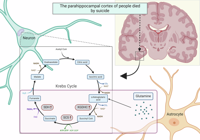
Our study, conducted with postmortem brain samples provided by the Semmelweis University Brain Tissue Bank, founded by the Prof. Miklós Palkovits, explored changes in energy-related processes in the PHC. We analyzed brain tissue from individuals who died by suicide and compared it with samples from individuals who died of other causes. The results highlight fascinating and significant molecular differences that could help us understand the biology of suicide.
Our study identified elevated levels of specific mitochondrial proteins in the PHC of individuals who died by suicide. These proteins are part of the tricarboxylic acid (TCA) cycle, a critical energy production pathway. Notably, these changes were absent in other brain regions, highlighting the PHC's unique role. Enzymes driving the TCA cycle are located in mitochondria cellular structures responsible for energy production, particularly in energy-demanding organs like the brain. Disruptions in mitochondrial function are linked to many mental health conditions, including depression. By investigating mitochondrial activity in the PHC, our research provided information on how energy production might change in individuals who died by suicide. The data suggest a phenomenon called glutaminolysis—a process where the brain converts glutamine, an amino acid, into energy, is increased in relation to suicide, signalling heightened neuronal activity in the PHC. While energy production is vital for brain function, excessive activity in areas like the PHC could potentially contribute to the thought patterns and emotional dysregulation associated with suicide.
Many individuals who die by suicide also suffer from major depressive disorder (MDD). Depression is known to impair mitochondrial function, but our findings suggest that some brain regions, like the PHC, might respond differently. Instead of reduced energy production, there seems to be a surge in energy in the PHC, potentially fueling overactivity that correlates with suicidal behavior. This research not only provides a molecular basis for understanding suicidal behavior but also opens the door for new interventions. By targeting the PHC or regulating mitochondrial activity, we might develop therapies that reduce the risk of suicide. Moreover, these findings underscore the importance of studying specific brain regions rather than generalizing across the entire brain. Such detailed approaches could yield more effective and personalized treatments.
While this study is a step forward, there’s still much to learn. Future research will explore how these metabolic changes in the PHC interact with other brain regions involved in depression and suicide. Understanding these networks could help unravel the complex biology behind suicide, ultimately guiding prevention efforts.
Follow the Topic
-
Translational Psychiatry
This journal focuses on papers that directly study psychiatric disorders and bring new discovery into clinical practice.

Your space to connect: The Psychedelics Hub
A new Communities’ space to connect, collaborate, and explore research on Psychotherapy, Clinical Psychology, and Neuroscience!
Continue reading announcementRelated Collections
With Collections, you can get published faster and increase your visibility.
From mechanism to intervention: translational psychiatry of childhood maltreatment
Publishing Model: Open Access
Deadline: Jun 30, 2026
Moving towards mechanism, causality and novel therapeutic interventions in translational psychiatry: focus on the microbiome-gut-brain axis
Publishing Model: Open Access
Deadline: Nov 15, 2026

Please sign in or register for FREE
If you are a registered user on Research Communities by Springer Nature, please sign in