Embrace Changes, Always

This is the first time our work is selected as the Featured Research in Journal of Translational Medicine. We are deeply honored. And here, I, the corresponding author, would like to share one of my untold stories behind this research.

Published in Biomedical Research

Embrace Changes, Always
Like

Share this post

Choose a social network to share with, or copy the URL to share elsewhere

This is a representation of how your post may appear on social media. The actual post will vary between social networks

Explore the Research

BioMed Central
BioMed Central BioMed Central

Decoding per- and polyfluoroalkyl substances (PFAS) in hepatocellular carcinoma: a multi-omics and computational toxicology approach - Journal of Translational Medicine

Background Per- and polyfluoroalkyl substances (PFAS), particularly perfluorooctanoic acid (PFOA) and perfluorooctane sulfonate (PFOS), are synthetic chemicals known for their widespread use and environmental persistence. These compounds have been increasingly linked to hepatotoxicity and the development of hepatocellular carcinoma (HCC). However, the molecular mechanisms by which PFAS contribute to HCC remain underexplored. Methods This study employs a multi-omics approach that combines network toxicology, integrated machine learning, single-cell RNA sequencing, spatial transcriptomics, experimental validation, and molecular docking simulations to uncover the mechanisms through which PFAS exposure drives HCC. We analyzed publicly available transcriptomic data from several HCC cohorts and used differential gene expression analysis to identify targets associated with both PFAS exposure and HCC. We constructed a protein–protein interaction (PPI) network and a survival risk model, the PFAS-related HCC signature (PFASRHSig), based on integrated machine learning to identify prognostic biomarkers, with the goal of identifying core targets of PFAS in HCC progression and prognosis. RT-qPCR and immunohistochemical (IHC) staining were used to validate the expression levels of the targets in both tumor and normal tissues. Molecular docking simulations were conducted to assess the binding affinities between PFAS compounds and selected target proteins. Results Functional enrichment studies revealed that PFAS targets were associated with metabolic signaling pathways, which are actively involved in lipid, glucose, drug metabolism, etc. Through integrated machine learning and PPI network analysis, we identified six genes, APOA1, ESR1, IGF1, PPARGC1A, SERPINE1, and PON1, that serve as core targets of PFAS in both HCC progression and prognosis. These targets were further validated via bulk RNA-seq, single-cell RNA-seq, and spatial transcriptomics, which revealed differential expression patterns across various cell types in the HCC tumor microenvironment. The results of RT-qPCR and IHC staining were consistent with the in silico findings. Molecular docking simulations revealed strong binding affinities between PFAS compounds and these core targets, supporting their potential roles in PFAS-induced hepatocarcinogenesis. Conclusions Our study highlights key molecular targets and pathways involved in PFAS-induced liver carcinogenesis and proposes a robust survival risk model (PFASRHSig) for HCC. These findings provide new insights into PFAS toxicity mechanisms and offer potential therapeutic targets for mitigating the health risks associated with PFAS exposure. Collectively, our findings help in advancing clinical applications by providing insights into disease mechanisms and potential therapeutic interventions.

I joined a research team in my freshman year in Wenzhou Medical University that  focused on bioinformatic analysis regarding breast cancer. The PI had big ambitions, as she spoke often of publishing groundbreaking papers and making a name in medical science. What she lacked, however, was the ability to turn any of those dreams into reality.

For two long years, she pushed us to satisfy her endless curiosity. Our own academic interests? Irrelevant. Our training needs? Ignored. She had us running analyses and generating data, all to support her ever-shifting, half-baked hypotheses.

Despite claiming herself as a mentor in computational biology, she could barely give any constructive suggestions and guidance. I had to spend my own money on R programming courses because the guidance I needed simply didn’t exist in her team. As for experimental design, she once had us perform T cell–related studies on nude mice (Balb/c), an act that defied scientific logic.

Still, I hesitated to leave. I feared that switching labs would “break the continuity” of my research training, as if there was anything coherent about the past two years.

Then the final straw came: one day during the summer holiday, after weeks of late nights spent in the lab, she said something cruel in a meeting, dismissive, cutting, and entirely uncalled for. That was it. I said goodbye.

After I left, I found a new mentor, who has real vision, solid expertise, and the real ability to lead a big team. I also met brilliant collaborators from across the country (e.g., Shanghai Jiao tong University Renji Hospital, BIOPIC, ...), who are thoughtful, capable, and passionate regarding their insights. For the first time, I felt part of a real scientific community, not just a cog in someone else's vanity project.

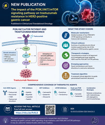
Then came the milestone that I finally got my first paper accepted. It was published in Journal of Translational Medicine. That achievement would have been unimaginable had I remained trapped in that dysfunctional lab. Stepping away didn’t just spare me further frustration, instead, it led me to a new environment filled with clarity, rigor, and collaboration.

That first publication was just the beginning. Encouraged and inspired by those around me, I went on to publish my second study, which, to my surprise, was selected as a Featured Research article. Yes, again in Journal of Translational Medicine

Now, as the first or corresponding author, I’ve published seven papers with a cumulative impact factor exceeding 50.  And more importantly, I love what I am doing now, deeply.

None of these would have happened if I hadn’t found the courage to leave the wrong place.

Embrace changes.

Always.

Please sign in or register for FREE

If you are a registered user on Research Communities by Springer Nature, please sign in

Follow the Topic

Biomedical Research
Life Sciences > Health Sciences > Biomedical Research

Related Collections

With collections, you can get published faster and increase your visibility.

Regeneration and Remodeling after Brain Injury: Implications for Stem Cell Therapy

Brain injury/damage, including brain trauma or stroke, has a high morbidity and mortality, and is often accompanied by severe neurological deficits post injury. Despite advances in therapeutics targeting brain repair and remodeling, positive clinical outcome is still a challenge and there continues to be a tremendous unmet clinical need for patients. Currently, transplantation of stem cells derived from several tissue sources has been recognized as very promising biologic-based therapy for regeneration and remodeling after brain injury, for instance, neural stem cells (NSCs), mesenchymal stem cells (MSCs) and induced pluripotent stem cells (iPS cells).

Upon brain injury, oxidative stress and inflammatory response are two early events in the biological cascade following brain injury. These two events are work synergistically and directly trigger the process of autophagy. Appropriate autophagy would contribute to brain recovery by reducing oxidative stress and inflammatory responses, whereas autophagy dysfunction exacerbates brain injury. Various studies have demonstrated that stem cells and their regenerative properties could impact tissue damage following brain injury in the following aspects: cellular replacement, secreting neurotrophic factors, and mitochondrial transfer. This collection aims to discuss the recent progress on stem cell therapy for brain injury, specifically studies elucidating the mechanisms in terms of oxidative stress, mitochondrion dysfunction and autophagy.

The collection welcomes all original and review articles on topics such as:

-Stem cell therapy for brain repair after brain injury/damage, including brain trauma or stroke;

-Underlying mechanisms upon stem cell replacement for brain injury, including cellular/tissue regeneration/remodeling, oxidative stress, inflammatory response and autophagy;

-Immune-response problems following stem cell therapy for brain injury;

-Mitochondrial role after stem cell therapy for brain injury;

-Detection of stem cell therapy for brain injury using imaging techniques, i.e. brain magnetic resonance;

Application of new tools, techniques, or models for studying stem cell viability or survival after implantation, such as organoids.

All submissions in this collection undergo the journal’s standard peer review process. Similarly, all manuscripts authored by a Guest Editor(s) are handled by the Editor-in-Chief. As an open access publication, this journal levies an article processing fee (details here). We recognize that many key stakeholders may not have access to such resources and are committed to supporting participation in this issue wherever resources are a barrier. For more information about what support may be available, please visit OA funding and support, or email OAfundingpolicy@springernature.com or the Editor-in-Chief.

Publishing Model: Open Access

Deadline: Ongoing

Microbiome in the first 1000 days of life

This collection aims to highlight recent advances in microbiome research during early life, focusing on its dynamics, impact on immune development, metabolism, and neurodevelopment, as well as potential interventions to promote a healthy microbiome. We welcome original research and review articles covering topics such as: 1. The role of the maternal microbiome in shaping infant microbiome development. 2. The impact of pregnancy complications on early-life microbiome composition. 3. Role of the human milk microbiome on infant’s health. 4. The influence of antibiotics and other early-life exposures on microbiome maturation. 5. The gut-brain axis: microbiome interactions and neurodevelopmental outcomes. 6. Interventions to promote a healthy microbiome, including probiotics, prebiotics, and dietary modifications

All submissions in this collection undergo the journal’s standard peer review process. Similarly, all manuscripts authored by a Guest Editor(s) will be handled by the Editor-in-Chief. As an open access publication, this journal levies an article processing fee (details here). We recognize that many key stakeholders may not have access to such resources and are committed to supporting participation in this issue wherever resources are a barrier. For more information about what support may be available, please visit OA funding and support, or email OAfundingpolicy@springernature.com or the Editor-in-Chief.

This Collection supports and amplifies research related to SDG 3, Good Health and Well-Being.

Publishing Model: Open Access

Deadline: Dec 17, 2025