From “Unnecessary” to “Just Right”: A GTPase Cracks the Rhoptry Secretion Secret in Apicomplexa
Published in Microbiology and Cell & Molecular Biology

Act I – What we knew about rhoptry secretion
Obligate intracellular parasites of the phylum Apicomplexa have only seconds to break into a host cell and take control. To do this, they carry a set of specialized “weapons” at their anterior end employed to rapidly secrete virulence factors essential for host-cell invasion and hijacking. The most dramatic of these elements are the rhoptries: pear-shaped organelles that inject proteins into the host cell, crossing not only the parasite’s plasma membrane but also that of the host.
In apicomplexan parasites such as Toxoplasma and Plasmodium, rhoptries are essential for invasion. Yet exactly how these organelles discharge their effectors at the right place and the right time is unclear—a gap that is both biologically critical and scientifically intriguing. We first discovered a complex of proteins (“Nds”) that is conserved not only throughout Apicomplexa but also in their close relatives, the ciliates, and found that they are essential for rhoptry discharge. We also identified a secretion machinery comprising an apical vesicle (AV) and a rhoptry secretory apparatus (RSA) that connects the AV to the parasite plasma membrane. Rhoptries are distinctive among eukaryotic secretory organelles in being constitutively docked to the parasite plasma membrane via the AV and RSA (Fig. 1)—like molecular syringes poised to fire, with host-cell contact serving as the discharge signal. Free-living ciliates use a similar strategy, with secretory organelles pre-docked at the plasma membrane and activated by environmental cues. This raises fundamental regulatory challenges: how do these systems remain primed without firing, and how are they activated at precisely the right time and place?
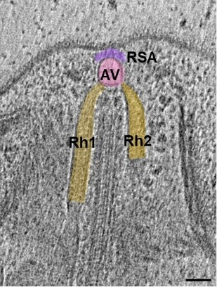
Figure 1: Two-dimensional slice from a tomogram of the apical complex of T. gondii displaying two rhoptries (brown), the AV (pink) and the RSA (purple). Scale bar: 50 nm.
In short: we knew rhoptries had to inject effectors, we knew roughly where and when, and we knew some of the cast. What we didn’t know was how the system is gated or prevented from misfiring.
Act II – Presenting our star: the Toxoplasma Nd-GTPase
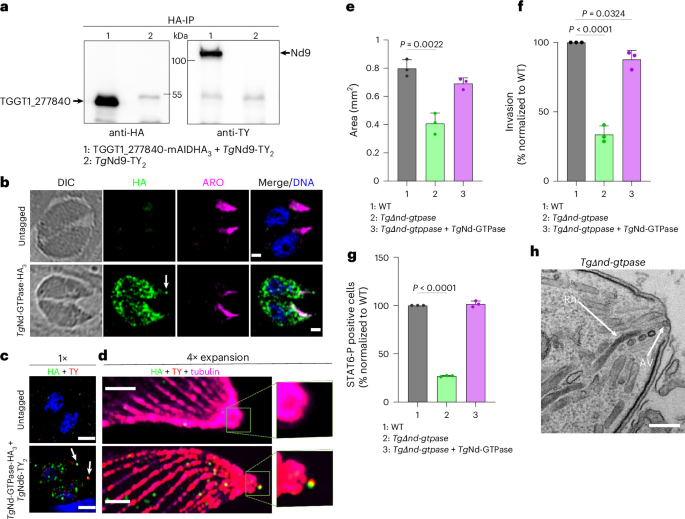
Every good story needs an unlikely protagonist. Ours is a previously uncharacterized small GTPase belonging to the Nd complex and beautifully presents at the apex of the parasite (Fig. 2).
GTPases are famous for being molecular switches regulating plenty of cellular processes, but this one didn’t set off any alarm bells: when we first looked at it, it appeared not to be essential for invasion or growth under standard in vitro conditions, showing only a half defect in rhoptry secretion. Because it was “dispensable” for parasite survival, it seemed a less exciting protein than initially anticipated. Thus, for a while, this GTPase was quietly moved to the bottom of our to-do list.
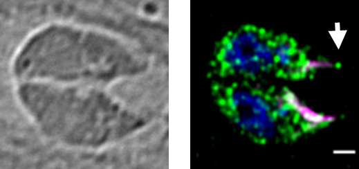
Figure 2: IFAs of intracellular tachyzoites endogenously expressing Nd-GTPase (green).
The arrow in indicates TgNd-GTPase in an apical punctum. Rhoptries (pink); nucleus (blue).
But here’s where the plot thickened. This GTPase caught our attention when we realized that it was conserved within the alveolates (including the parasites’ ciliate cousins) and does not fit neatly into any traditional family of small GTPases, such as Ras or Rab, suggesting an Alveolata-specific function. On top of that, because the protein was not essential, we did not end up with a dead parasite simply telling us, “This protein matters.” Instead, we had a living, yet secretion-deficient, parasite that whispered, “This protein fine-tunes the system.” So we started asking mechanistic questions: who are the partners in crime of this GTPase? Is it always present at the secretion site? How does it change the behavior of the rhoptry machinery? Biochemistry and genetics could tell us that the GTPase influences rhoptry discharge, that the GTP-bound form is the active form, that it interacts directly with the essential Nd9 protein, but it couldn’t show us whether this defect was associated to structural changes at the apex of the mutant parasite. For that, we called in our trusted detectives: the cryo-electron tomography (cryo-ET) experts.
Cryo-ET allowed us to visualize the entire machinery in 3D, in situ and in a near-native state at nanometer resolution. In wild-type Toxoplasma, we saw the familiar cast: rhoptries, an AV, and the beautiful eightfold-symmetric architecture of the RSA. Everything looked ordered, restrained, and ready to fire (when the right signal arrived). Our earlier RSA structures were not sufficiently resolved to assign individual Nd components. Through a structural biology tour de force that substantially improve the RSA resolution, combined with AI-assisted integrative structural modeling, we were finally able to position the GTPase in complex with Nd9 and NdP1 within the RSA. The GTPase occupies a central position within the RSA (Fig. 3), ideally placed to regulate the machinery. In the GTPase mutant, the picture changed. The overall architecture was still there, but the RSA-AV interface and RSA conformation were perturbed in a subset of parasites. Strikingly, this dual phenotype led to two scenarios: one in which the AV-RSA interface becomes leaky, potentially permitting initiation of secretion; and another in which the RSA collapses and locks the interface, likely preventing secretion altogether.
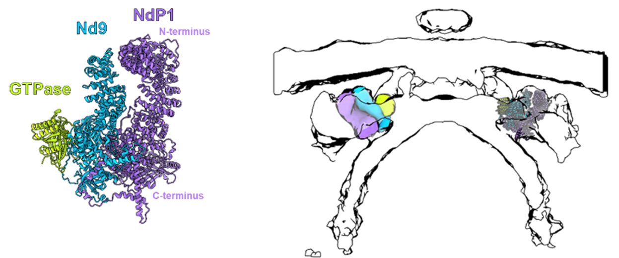
Figure 3: Left, Alphafold 3.0 predicted structure of Toxoplasma Nd-GTPase/Nd9/NdP1 complex.
Right, Structural fitting of the complex within the RSA (side view).
Suddenly, the GTPase was no longer just a genetic hit; it had a structural footprint. The cryo-ET data gave us the first glimpse of how a single regulator can shape the conformation and behavior of the RSA.
Act III – The rise of our co-star: a Tetrahymena Nd-GTPase
Every hero needs a co-star, and ours turned out to be the free-living ciliate Tetrahymena thermophila. Like Toxoplasma, Tetrahymena belongs to the Alveolata and has secretory organelles called mucocysts that are broadly analogous to rhoptries. It also carries a homolog of our supposedly “dispensable” Toxoplasma Nd-GTPase. When we began to examine this protein in Tetrahymena, our story took a fascinating turn.
This GTPase is also part of the Nd complex in Tetrahymena and partially responsible for secretion upon stimulation. In Tetrahymena, losing this GTPase had a remarkable effect: the cells began to spontaneously secrete the contents of their mucocysts into the medium, without any stimulation. No control! Just “go with the flow,” and mucocyst proteins end up in the medium.
This mutant gave us the first real clue to what the GTPase might be doing. Rather than being a passive bystander, it acts as a brake on secretion: in its absence, the system defaults to leaky secretion mode. This gatekeeping function depends on the GTP-bound form in association with two partners in crime: Nd9 and NdP1 proteins. This pointed toward a potential role of the Alveolata-specific Nd-GTPase/Nd9/NdP1 trio in controlling membrane fusion or the final steps of exocytosis. Et voilà, the trio is conveniently located within the RSA structure to do this job (go to the paper if you want to know more!).
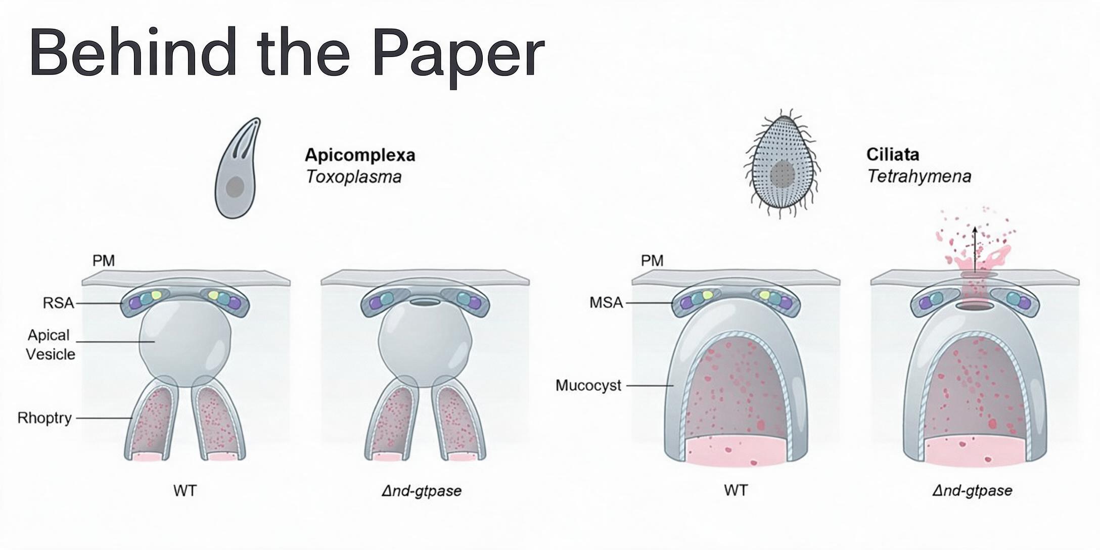
But then came the obvious, slightly annoying question: if the GTPase acts as a brake in Tetrahymena, why don’t we see the same runaway secretion in the Toxoplasma mutant? This brings us to another central feature of apicomplexan parasites: the AV (absent in ciliates!). Its position between the rhoptry and RSA might represent a physical barrier that prevents rhoptry leakage (Fig. 4). However, whether this is its only function of the mysterious AV remains to be determined.
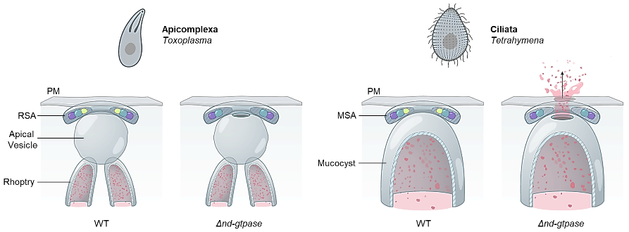
Figure 4: Model for Nd-GTPase function in Apicomplexa and Ciliata.
RSA: rhoptry secretory apparatus; MSA: mucocyst secretory apparatus; PM: plasma membrane;
green circle: Nd-GTPase; blue circle: Nd9; purple circle: NdP1.
Long story short: building on a protein that initially looked dispensable, we obtained the first mechanistic view of how this unusual alveolate secretion machinery is gated. The Nd-GTPase/Nd9/NdP1 complex helps stabilize a pre-secretion state and prevents uncontrolled exocytosis; upon stimulation, the gate must be released so that the machinery can rearrange and secretion can proceed. In Apicomplexa, this secretory system emerges as a regulatory hub that confers not only precision but also versatility, with the capacity to integrate additional elements such as the AV that might act as extra checkpoints and reinforce the regulatory mechanism required for host-cell invasion. Sometimes the most revealing proteins are not the ones whose deletion kills the cell outright. Sometimes, keeping the system alive but slightly broken is exactly what allows you to see how it works. From “dispensable” to gatekeeper, our initially underestimated GTPase gave us a way into the black box of alveolate exocytosis.

Follow the Topic
-
Nature Microbiology
An online-only monthly journal interested in all aspects of microorganisms, be it their evolution, physiology and cell biology; their interactions with each other, with a host or with an environment; or their societal significance.