How E. coli renews its outer membrane when growth slows
Published in Microbiology and Cell & Molecular Biology







The outer membrane is a defining feature of Gram-negative bacteria. It forms a formidable permeability barrier that protects the cell from toxic compounds, many antibiotics and components of the immune system. It also contributes directly to the strength and stability of the cell envelope. Both functions depend on lipopolysaccharide (LPS), the main component of the membrane’s outer leaflet. LPS molecules form a densely packed, highly ordered layer. Strong interactions between neighbouring molecules make this layer remarkably robust and restrict the movement of LPS after membrane insertion. Unlike the lipid components of a conventional fluid membrane, LPS therefore diffuses only very slowly across the cell surface.
This creates a fundamental problem for membrane renewal and adaptation which bacteria use to resist peptide antibiotics like polymyxin B and colistin. During rapid growth, the cell adds new material as its surface expands. Pre-existing material is then progressively diluted through elongation and division. But bacteria in most environmental and pathogenically relevant settings spend long periods growing slowly or not dividing at all. Under these conditions, surface expansion can no longer provide an efficient route for replacing existing LPS. How does the LPS content of the outer membrane still undergo adaptation when growth slows?
Following new LPS into the outer membrane
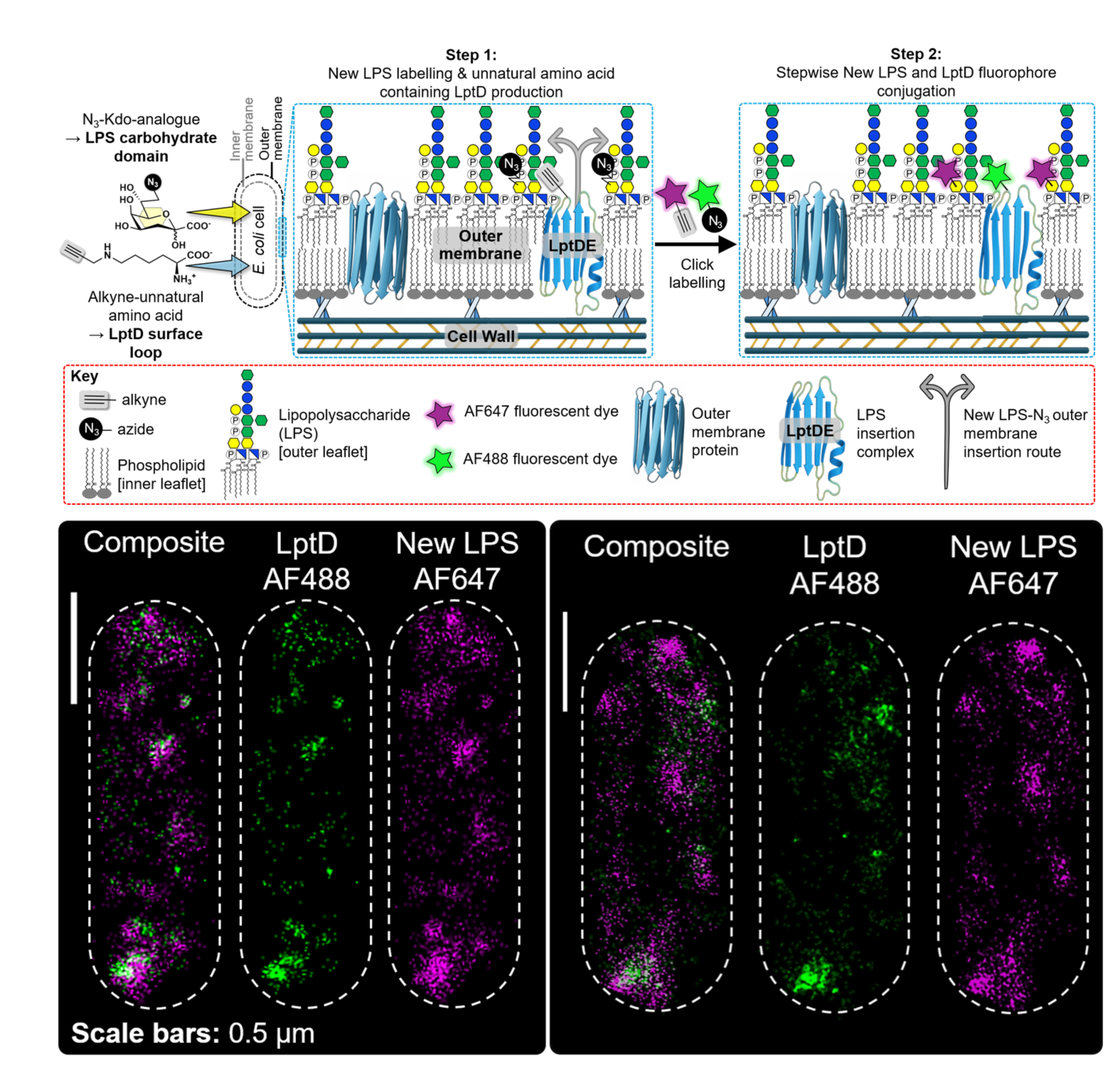
We first needed to find where newly synthesised LPS enters the cell surface. LPS is transported across the cell envelope by the Lpt machinery. At the outer membrane, the LptDE complex performs the final step and inserts LPS into the outer leaflet. To follow this process, we combined two complementary forms of bioorthogonal labelling to enable visualisation of these outer membrane components. Genetic
code expansion allowed us to attach a fluorescent dye to LptD at a defined site. Metabolic labelling was used to mark newly synthesised LPS with a second dye. Their combined application allowed us to visualise both the delivery machinery and its newly delivered cargo in the same cell (Figure 1). Two-colour super-resolution microscopy then allowed us to compare their positions at nanometre-scale resolution.
Both LptD and new LPS appeared as discrete puncta distributed across the outer
membrane. The two signals were strongly correlated over short distances. This was consistent with LptD-associated regions marking sites of LPS delivery. Unlike outer-membrane protein insertion, these sites were distributed across the entire cell surface. The close association also suggested that new LPS did not diffuse far immediately after its insertion.
Finding the insertion sites raised the next question. What happened to LPS after its insertion?
Colour coding different LPS vintages
To answer this, we needed to distinguish LPS of different vintages in the outer membrane of daughter cells through bacterial generations. We used multiple versions of chemically functionalised Kdo, a sugar in the conserved inner core carbohydrate region of LPS. Bacteria incorporated these Kdo analogues into LPS. Each analogue added a small, chemically unique handle that could later be “clicked” to a specific fluorescent dye depending on the nature of the chemical handle on the functionalised Kdo analogue (Figure 2).
By switching from one Kdo analogue to another during pulse–chase experiments, we labelled “pre-existing” LPS that was already present separately from “new” LPS inserted thereafter. In effect, we could colour code these different vintages of LPS and follow both populations in subsequent bacterial generations.
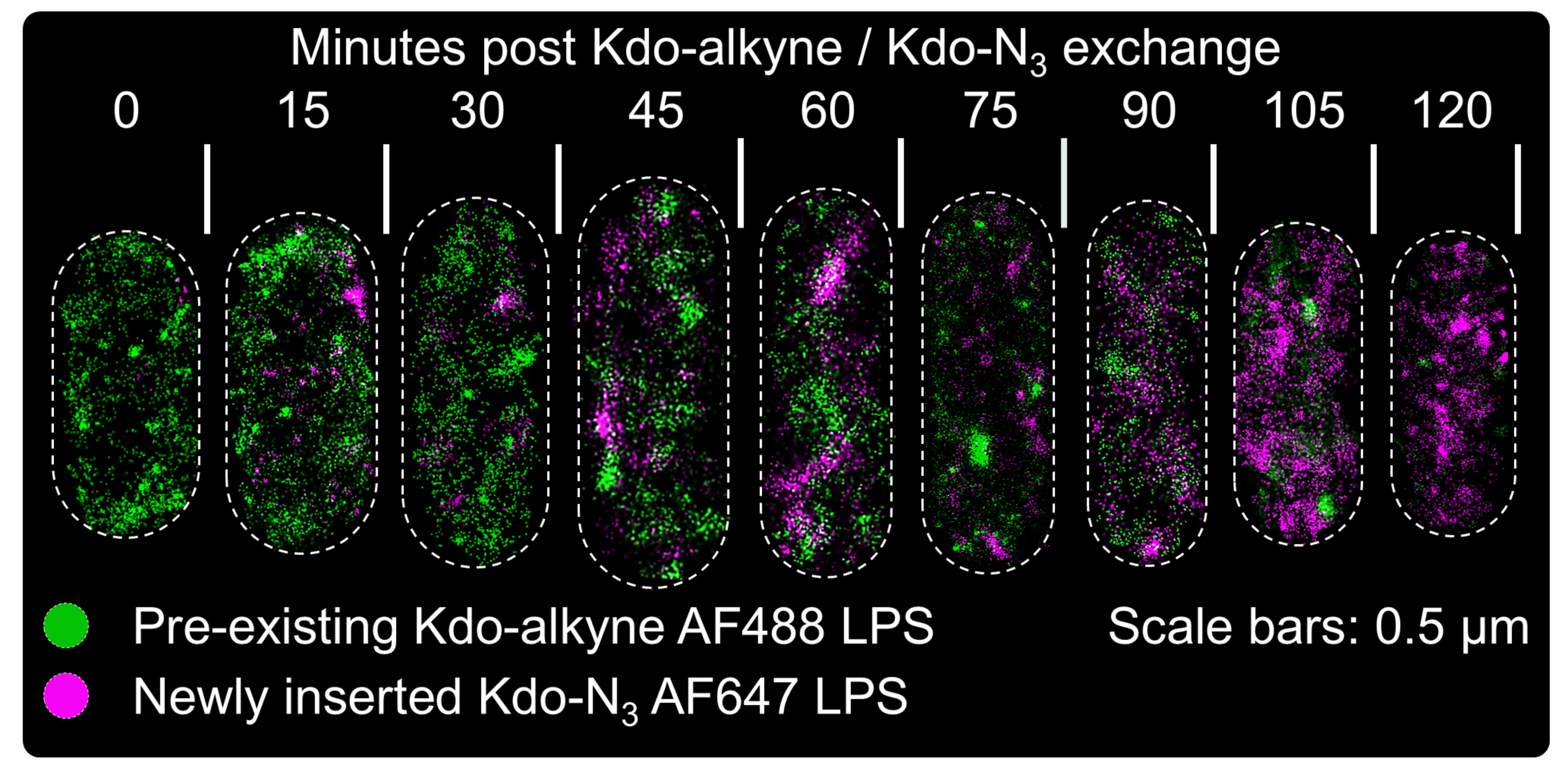
The results were striking. Newly inserted LPS accumulated in discrete patches that remained largely separate from the pre-existing population. Even as more new LPS entered the membrane, there was remarkably little large-scale mixing between the two vintages (Figure 2).
These observations led us to propose a new insertion-trapping model to describe LPS organisation. New LPS enters at discrete sites across the cell surface. Its lateral diffusion is so restricted that the molecules remain close to where they are inserted. Successive insertion events therefore create persistent LPS-rich patches. The organisation of the membrane is established during insertion and then kinetically trapped by the severely limited movement of LPS.
Where was the pre-existing LPS going?
The most unexpected result in our study emerged as cultures entered stationary phase.
As expected, cell elongation and division slowed markedly upon entry into stationary growth phase. Yet new LPS continued to enter the outer membrane at the same rate as was observed when cell elongation and division were occurring rapidly during exponential growth. At the same time, the pre-existing LPS population continued to decrease. Its loss was significantly greater than could be explained by growth-dependent dilution alone.
The sustained rate of LPS turnover despite a slowing in cell growth led us to conclude that the two processes, LPS insertion and cell elongation, were becoming uncoupled. If pre-existing LPS was disappearing from the cell surface in the absence of cell growth, where was it going?
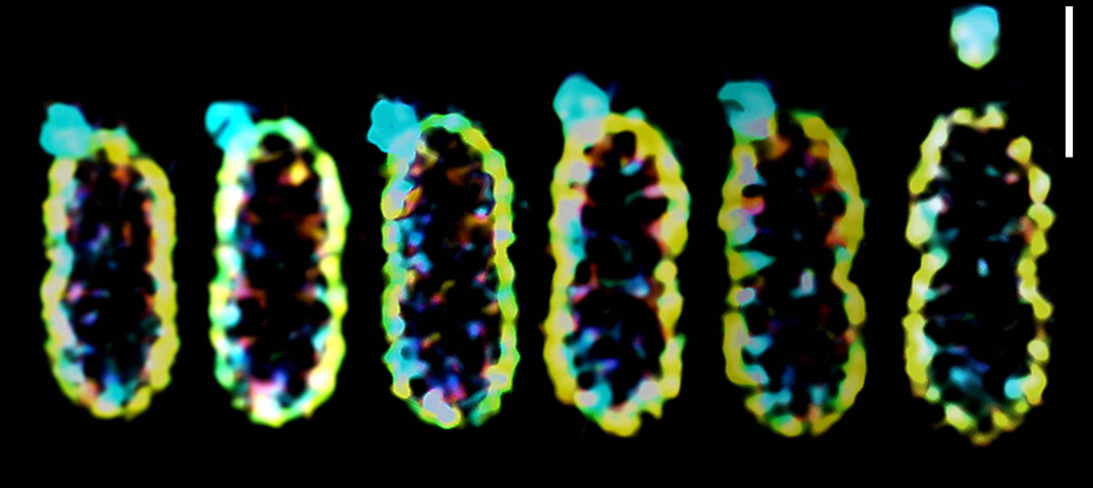
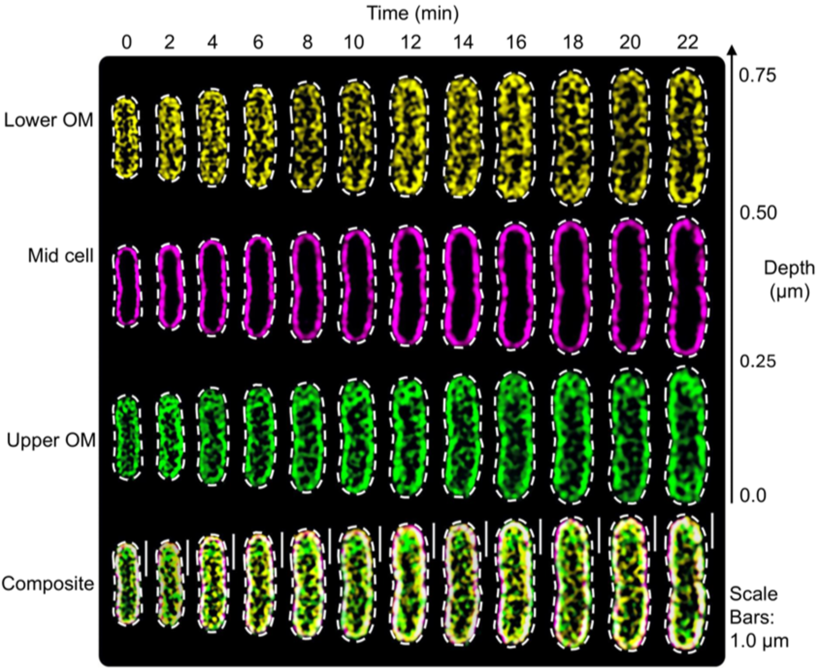
We first considered whether it was moving to another part of the membrane. Live-cell 3D super-resolution microscopy showed no progressive accumulation at the cell poles, as observed previously for binary partitioning of outer membrane proteins (Figure 3). We also considered whether the cell was internalising and recycling pre-existing LPS. To test this, we fractionated a population of E. coli into their major cellular compartments and measured where the labelled LPS was found. There was no evidence that old LPS or its respective components were accumulating inside the cell.
|
Figure 3. Pre-existing LPS remains distributed across the outer membrane and is not recycled inside the cell. Live-cell 3D super-resolution images show optical sections through the lower outer membrane, the middle of the cell and the upper outer membrane over 22 minutes. The labelled LPS remains as patchy regions at the cell surface. There is no progressive accumulation at the cell poles or within the cell. Colours distinguish the different imaging planes. Dashed lines indicate cell boundaries. |
A route out through vesicles
A Gram-negative bacterium can naturally release a small portion of the outer membrane as an outer membrane vesicle (OMV). OMVs are widely studied as vehicles for secretion, cargo delivery and interactions with other bacteria and host cells. We theorised that they may also provide a direct physical route by which membrane material can leave the bacterial surface.
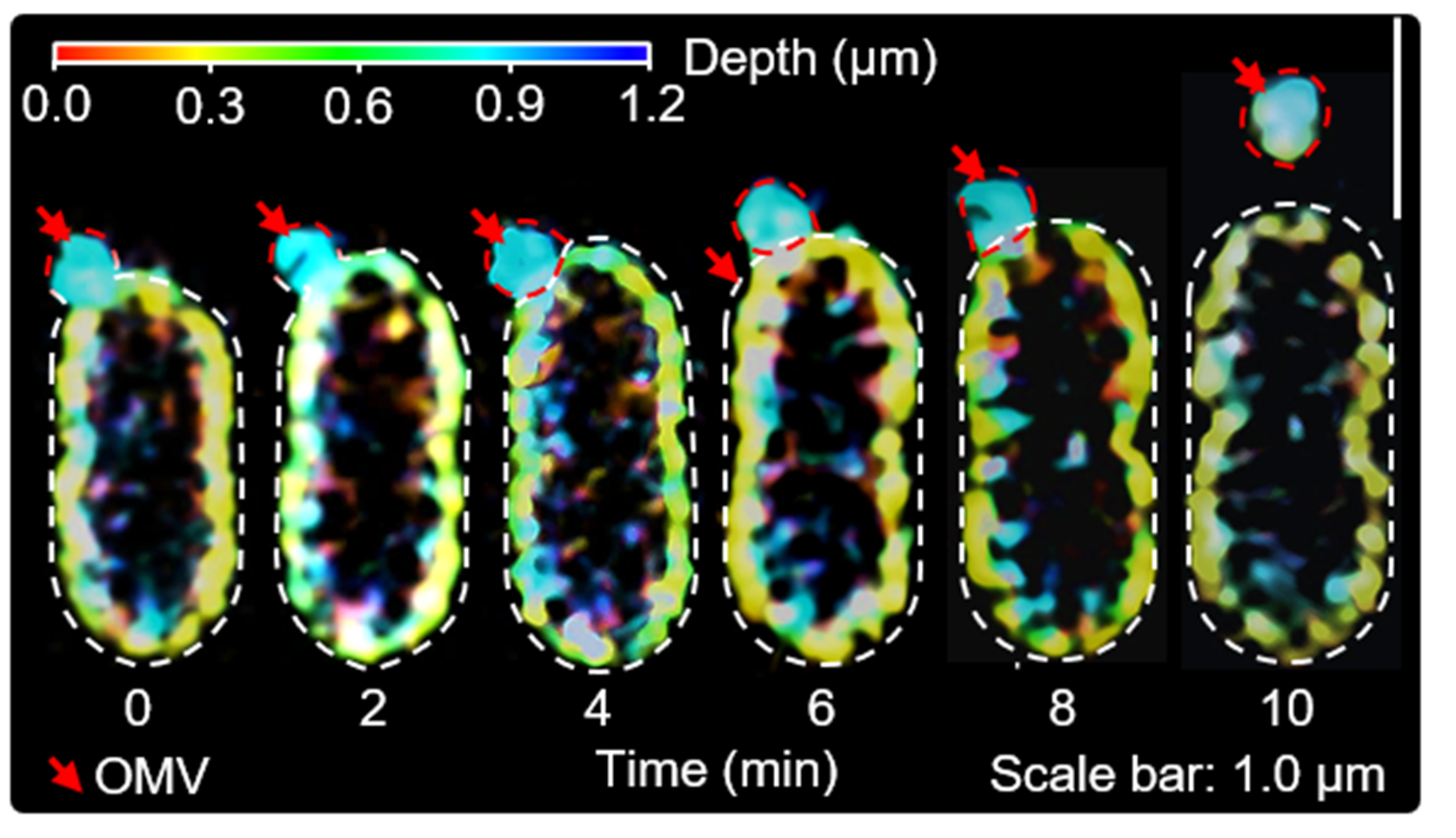
One of the most memorable observations in the project came during live-cell imaging. We were able to capture the deformation and outward extension of an LPS-rich region in the outer membrane, and it detaching as a discrete OMV (Figure 4). We had just demonstrated that OMV biogenesis could be visualised at the single cell level using super-resolution imaging.
|
Figure 4. Watching an outer membrane vesicle. A depth-coloured live-cell 3D super-resolution time series shows an LPS-rich region of the outer membrane deforming, extending outwards and detaching as an outer membrane vesicle. Red arrows mark the developing vesicle. Colours represent imaging depth rather than different LPS populations. Dashed lines indicate the cell and vesicle boundaries. |
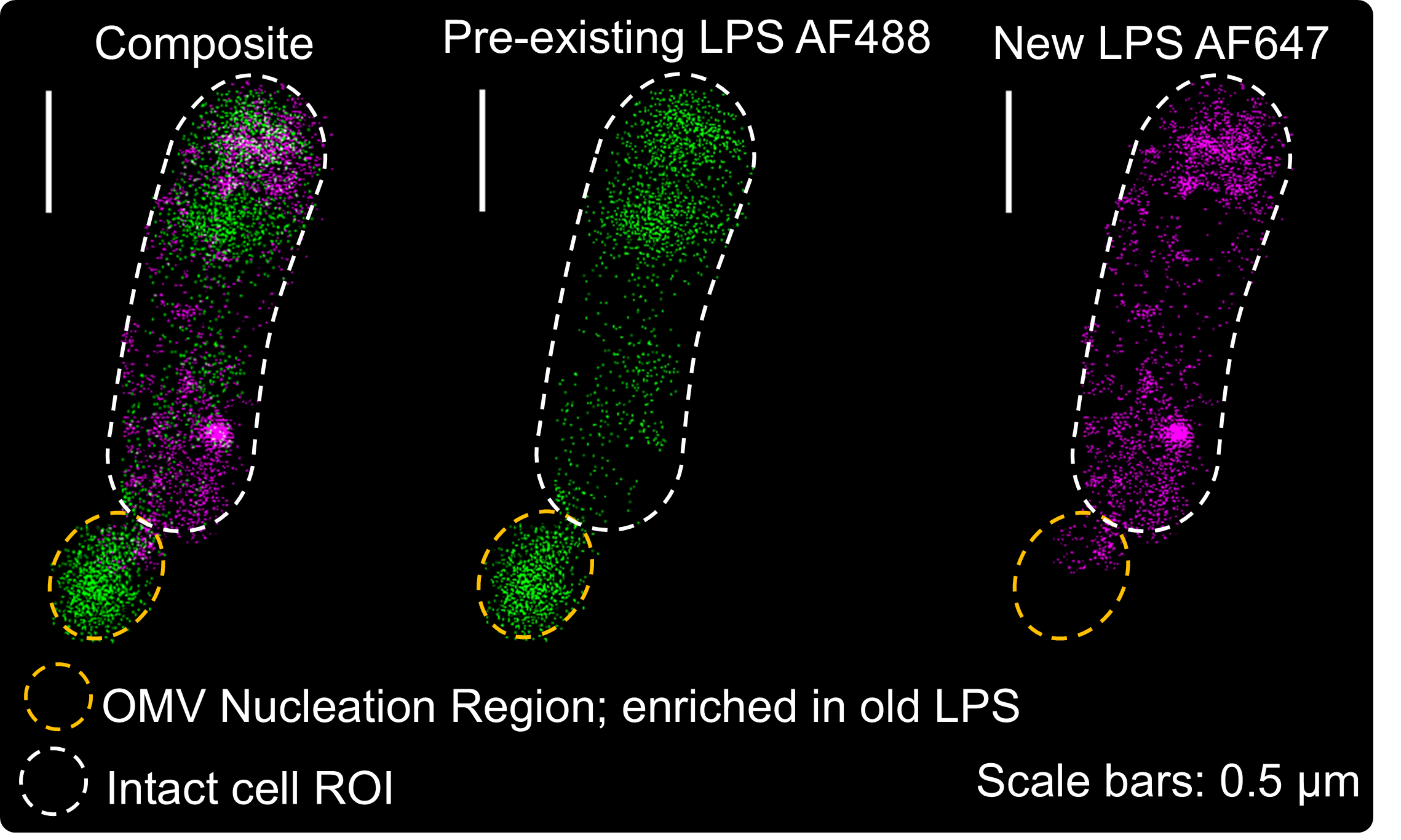
Two-colour dSTORM experiments done after labelling of pre-existing and new LPS showed that OMV-like protrusions were preferentially enriched in pre-existing LPS and relatively depleted in newly inserted LPS (Figure 5). Across the events we analysed, pre-existing LPS showed a median enrichment of approximately 4.2-fold within OMV regions compared with the surrounding outer membrane.
Population-level measurements supported these microscopy-based results. Labelled pre-existing LPS decreased in isolated outer membranes whilst simultaneously increasing in the OMV fraction. Nanoparticle tracking also showed increased vesicle production as cultures entered stationary phase where cell growth halts. Quantitative modelling indicated that OMV export could account for a substantial component of LPS loss beyond that expected from growth-dependent dilution.
Together, these observations identify OMV release as an important route for growth-independent LPS turnover, a previously unreported OMV function.
This does not mean that the cell recognises individual LPS molecules based on their vintage. Rather, preferential removal of pre-existing LPS could instead emerge from the heterogeneous spatial organisation and physical properties of the outer membrane. New LPS remains localised near insertion sites, while vesiculation disproportionately removes regions rich in pre-existing LPS, found at relatively greater distances from tightly pinned regions enriched in outer membrane proteins.
A new perspective on LPS organisation and turnover in the outer membrane
It is well-established that the spatial organisation of the outer membrane is heterogeneous, existing as a patchwork of LPS-rich and protein-rich regions or ‘clusters’. Outer membrane protein clusters can associate into a supramolecular network covering the entire bacterial cell surface. Interspersed within this network are LPS-rich clusters. What drives the formation of this spatial heterogeneity?
Phase separation has been proposed as one explanation for the coexistence of LPS-rich and protein-rich clusters in the outer membrane. Such a mechanism makes a clear prediction. Over time, these domains should coarsen. Smaller LPS or protein regions should disappear or merge, while larger regions progressively grow.
We did not observe this behaviour for LPS. Newly inserted LPS patches increased in number, but their average size eventually plateaued. Pre-existing patches diminished without progressively merging into larger regions. Particle-based simulations sharpened this distinction. A coarse-grained outer membrane model that combined localised insertion, restricted lateral diffusion and OMV-mediated loss reproduced the persistent separation of new and pre-existing LPS without domain coarsening. A phase-separation model with the same overall insertion and turnover constraints behaved very differently. It predicted an increase in co-clustering and progressive domain growth.
The resulting picture is of a kinetically trapped, non-equilibrium outer membrane. Localised insertion establishes new LPS-rich regions while restricted lateral diffusion preserves that organisation. Vesiculation provides a route for pre-existing material to leave. Together, these processes allow the LPS composition of the membrane to remain dynamic even when growth-dependent membrane expansion and turnover is strongly limited. It remains to be determined whether growth-independent LPS turnover is a defining feature of Gram-negative bacteria, and how environmental stresses might alter this process. Regardless, the central message is clear - Gram-negative bacteria have the capacity to renew LPS composition in a growth-independent manner.

I am a chemical biologist at the University of York developing molecular, imaging and computational tools to reveal and ultimately control dynamic biological interfaces. My work spans Gram-negative bacterial outer membranes and cell-instructive biomaterials, united by a central question: how does spatial organisation at a biological surface determine its behaviour?
My bacterial research focuses on lipopolysaccharide (LPS), the principal component of the Gram-negative outer membrane, and outer membrane vesicles (OMVs). By developing novel, innovative selective metabolic and bioorthogonal labelling strategies, I have been able to chemically distinguish different generations and glycoforms of LPS and follow their organisation at single-cell and nanoscale resolution. This work revealed that interactions within the conserved Lipid A anchor restrict LPS mobility, while localised insertion, persistent segregation of pre-existing and newly inserted LPS, and OMV-mediated removal allow the outer membrane to renew even as bacterial growth slows. These discoveries led to a corresponding-author paper in Nature Communications and a first-author paper in The EMBO Journal.
I am now building on this research to understand how distinct LPS surface states influence OMV biogenesis and antimicrobial protection, with the longer-term aim of informing new outer-membrane-targeting strategies and OMV-based technologies. In parallel, I lead an application-driven project in the Spicer Group developing light-responsive, peptide-functionalised hydrogels that control neuronal adhesion and neurite outgrowth. Across both areas, my aim is to create chemical tools that move beyond observing biological organisation towards manipulating it with spatial and temporal precision.
Core Expertise
-
Chemical and analytical science: Multistep organic and carbohydrate synthesis; Fmoc solid-phase and flow-based peptide synthesis; peptide and protein modification; bioconjugation; bioorthogonal-probe development and live-cell labelling; advanced characterisation using NMR, LC–MS(/MS), analytical HPLC, fluorescence assays and gel-based methods; and light-responsive, peptide-functionalised biomaterials.
-
Cellular and microbiological systems: Gram-negative outer-membrane biology, LPS and OMVs; bacterial manipulation and recombinant protein production; extensive experience with model organisms and pathogenic isolates under Containment Level 2 and enhanced CL2 conditions; and eukaryotic cell biology spanning primary-neuron isolation and culture, mammalian cell culture, and primary and immortalised immune-cell models.
-
Imaging, biophysics and computation: advanced live-cell and super-resolution fluorescence microscopy, including multicolour dSTORM, 3D-SIM², FRAP and single-particle tracking; confocal and spinning-disk imaging; and bespoke MATLAB and Python pipelines for automated image quantification, spatial statistics, clustering, cross-correlation and mechanistic modelling.
Follow the Topic
-
Nature Communications
An open access, multidisciplinary journal dedicated to publishing high-quality research in all areas of the biological, health, physical, chemical and Earth sciences.

Related Collections
With Collections, you can get published faster and increase your visibility.
Women's Health
Publishing Model: Hybrid
Deadline: Ongoing
Tumor Microenvironment Crosstalk and Therapeutic Implications
Publishing Model: Hybrid
Deadline: Nov 02, 2026