Insights into the evolution of polymer crystals by 2D and 3D electron nanodiffraction imaging
Published in Materials




Why study polymers?
Plastic pollution is one of the most critical environmental challenges of our time. Although polymers are deeply embedded in modern life, we are yet to fully understand how their internal structures form, evolve, and ultimately influence their properties and degradation. Gaining this understanding is essential for designing better sustainable plastics and improving strategies for their recycling and disposal.
How did this project start and what is its main goal?
This project, initiated by Dr. Anastasiya Sedova and Prof. Boris Rybtchinski at the Weizmann Institute of Science, set out to investigate polylactic acid (PLA), one of the world’s most widely used compostable plastics. PLA is increasingly used in packaging, consumer products, textiles, biomedical materials, and 3D printing, making it a highly relevant system for both industry and sustainability research1.
The goal of this study was to understand how industrial thermal processing methods, specifically extrusion, injection molding, and annealing, affect the crystallization of PLA. These processing steps are widely used in plastics manufacturing and strongly influence the final mechanical properties of the material2. However, the relationship between processing conditions and nanoscale polymer structure has remained poorly understood.
Experimental approach
To mimic the industrial processing conditions for PLA, we used a mini-extruder and mini-injection molding machines suitable for processing small quantities of the polymer. The thermal treatment that followed these processes resulted in various crystalline phases and different degrees of crystallinity of PLA.
Using a combination of advanced electron nanodiffraction imaging (4D-STEM)3, atomic force microscopy (AFM), X-ray diffraction (XRD), and differential scanning calorimetry (DSC), we visualized the internal architecture of PLA in both two (2D) and three dimensions (3D) (Fig. 1). Remarkably, we were able to image highly beam-sensitive polymer crystals at nanometric resolution and to reveal how polymer chains organize inside crystalline domains known as lamellae.
About 4D-STEM and 4D-STEM tomography
The 4D-STEM technique3 was developed using a transmission electron microscope (TEM). This method uses a focused electron beam (probe) to scan the sample, also termed scanning transmission electron microscopy (STEM). By combining the STEM approach with a high-speed hybrid pixel array detector, we can collect an electron diffraction pattern at each x-y scan position. The number of diffraction patterns that can be acquired (data set) depends on the number of pixels chosen for image reconstruction. For example, 16,384 patterns are collected for a scan grid of 128x128, 262,144 patterns for a grid of 512x512, and so on. Scan grid size should be evaluated carefully, especially for beam-sensitive materials, such as polymers, since increasing the number of pixels can lead to overlapping areas and, consequently, additional radiation damage to the analyzed samples. A significant number of diffraction patterns allows information to be obtained from every pixel of the scanned sample with a nanometric resolution.
The 4D-STEM tomography4 is an extension of the 4D-STEM technique, in which the sample is tilted from 0° to ±60° angle in 3° increments, and a data set is being collected at each tilt. A complex computational approach is required for data analysis to transform the acquired electron diffraction data into frames that can be combined to create a 3D image.
Main results of this work
Our results show that thermal processing drives the formation of hierarchical crystalline structures composed of stacked lamellar crystals that grow together into highly ordered bundles. The polymer chains inside these lamellae are tilted rather than perfectly aligned, an important structural feature that influences crystal packing and ultimately affects the mechanical behavior of the material.
One of the most significant achievements of this project was the successful implementation of 3D nanodiffraction tomography for a polymeric system. Until now, this highly sophisticated imaging approach had primarily been demonstrated in biological samples4. By adapting it for polymers, we reconstructed the three-dimensional organization of PLA crystals and directly visualized interacting lamellar networks with unprecedented detail. This represents an important methodological breakthrough for polymer science.
Beyond the technical accomplishment, the findings provide new insight into how biodegradable plastics develop their internal structure during manufacturing. Because the arrangement of polymer chains determines properties such as strength, flexibility, and degradation behavior, understanding these nanoscale architectures is critical for the future design of sustainable materials. This study therefore advances not only our fundamental understanding of polymer crystallization, but also the broader effort to create plastics with improved performance and more predictable environmental fate.
On a methodological side, by combining high-resolution orientation mapping, chain-tilt analysis, and 3D nanodiffraction imaging, we established a new framework for studying polymer crystallization across multiple length scales.
This research was completed with the active collaboration of Dr. Lothar Houben, Dr. Shahar Seifer, and Dr. Sidney R. Cohen.
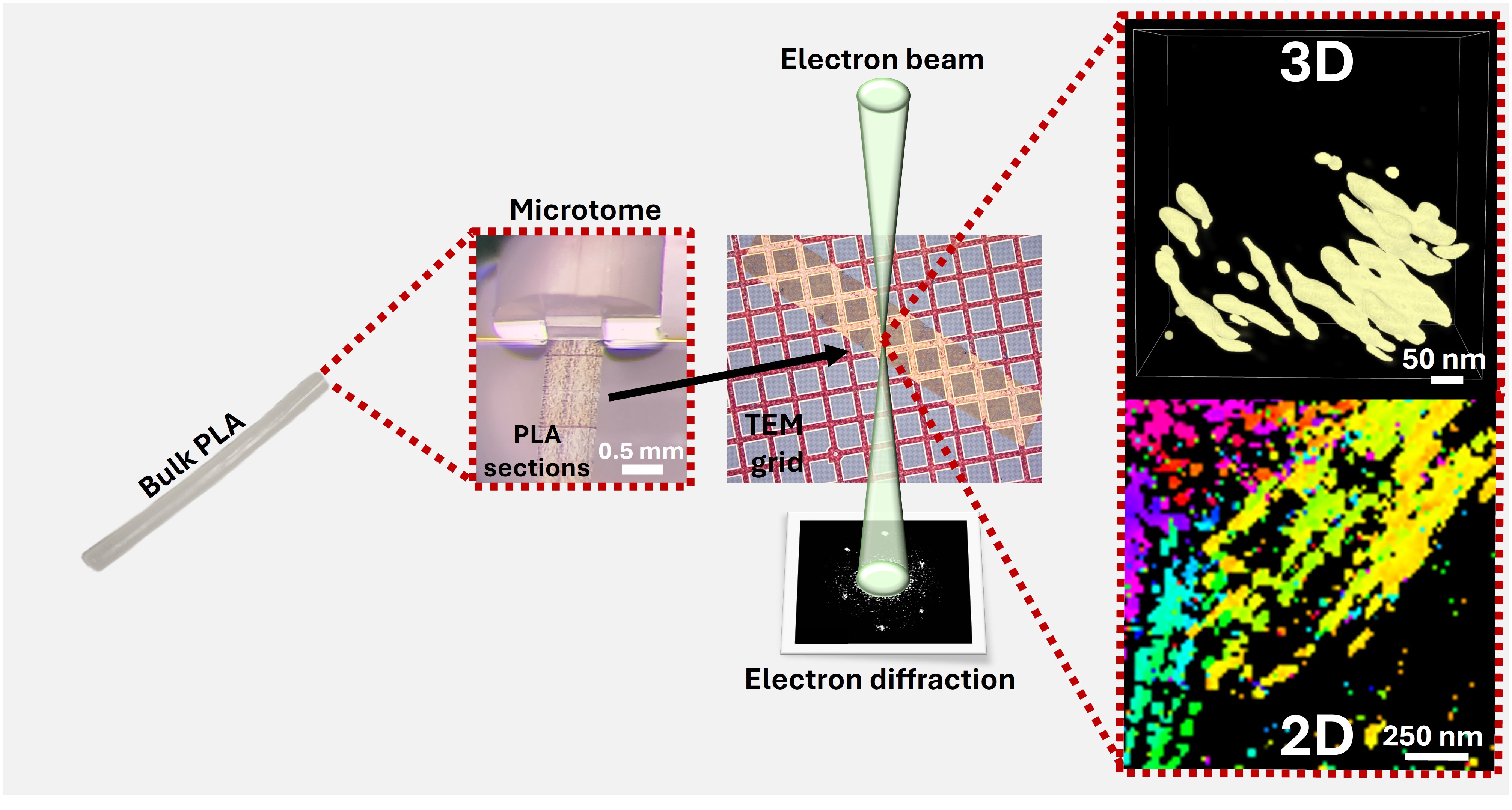
Fig. 1 Visualizes the preparation of thin sections from the bulk PLA using a microtome. These thin sections are then transferred to a TEM grid for analysis by 4D-STEM. Finally, the collected diffraction patterns are processed computationally to obtain the 2D and 3D images.
References:
- Sin, L. T. & Tueen, B. S. Polylactic acid: a practical guide for the processing, manufacturing, and applications of PLA. (William Andrew, 2019).
- Vlachopoulos, J. & Strutt, D. Polymer processing. Mater. Sci. Technol. 19, 1161–1169 (2003).
- Bustillo, K. C. et al. 4D-STEM of beam-sensitive materials. Acc. Chem. Res. 54, 2543–2551 (2021).
- Seifer, S., Kirchweger, P., Edel, K. M. & Elbaum, M. Optimizing contrast in automated 4d stem cryotomography. Microsc. Microanal. 30, 476–488 (2024).
Follow the Topic
-
Communications Materials
A selective open access journal from Nature Portfolio publishing high-quality research, reviews and commentary in all areas of materials science.

Related Collections
With Collections, you can get published faster and increase your visibility.
Orbitronics and unconventional spin-orbit torque for next-generation magnetic memory and spin logic devices
Publishing Model: Open Access
Deadline: Oct 31, 2026
Smart windows for sustainable buildings
Publishing Model: Open Access
Deadline: Oct 31, 2026