Investigating inflammasomes and cell death
Published in Protocols & Methods
Written by Rebecca Tweedell and Thirumala-Devi Kanneganti
The innate immune system acts as the first line of defense against infectious and sterile insults. Inflammasomes and cell death are key components of the innate immune system. While inflammasome-mediated innate immune activation and cell death are beneficial in the context of clearing pathogens and removing damaged cells, these processes can also have detrimental effects and lead to uncontrolled inflammation and immunopathology. Therefore, understanding inflammasome activation and cell death are critical for maintaining homeostasis and preventing disease. Methods to study these processes open the door for the identification of novel regulators and effectors in these pathways that can serve as therapeutic targets to improve the treatment of infectious, inflammatory, and metabolic diseases and cancers.
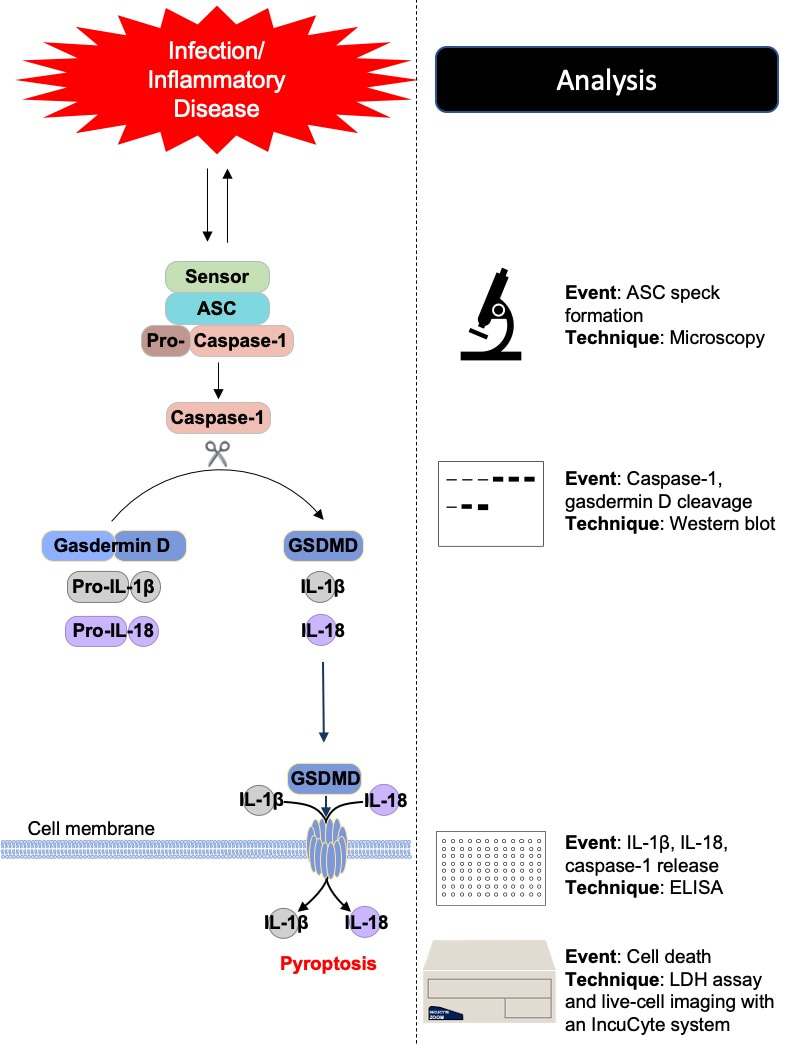
The inflammasome is a multiprotein complex that assembles in response to pathogen- or damage-associated molecular patterns. Inflammasomes are canonically composed of a sensor, the adaptor protein ASC, and the effector protein caspase-1. Inflammasomes mediate caspase-1 activation, which in turn cleaves its substrates, including pro–IL-1β, pro–IL-18, and gasdermin D. This cleavage frees the N-terminus of gasdermin D to form pores in the cell membrane, facilitating the release of matured IL-1β and IL-18 and executing a form of proinflammatory cell death called pyroptosis.
Inflammasome activation and subsequent cell death involve a number of steps that can each be monitored to provide a comprehensive picture of the kinetics, molecular details, and functional outcomes of the process. Researchers have historically focused on a single step or a small handful of steps to indicate whether inflammasome activation is occurring. However, this provides an incomplete snapshot of the biology, as completion of one step does not automatically guarantee completion of all steps. For example, the release of proinflammatory cytokines and execution of cell death downstream of caspase-1 activation can be blocked by a defect in gasdermin D or the action of an inhibitory protein from a pathogen. It is essential to monitor each step in the pathway to provide a comprehensive view of inflammasome activation and cell death and to identify roadblocks in the process.
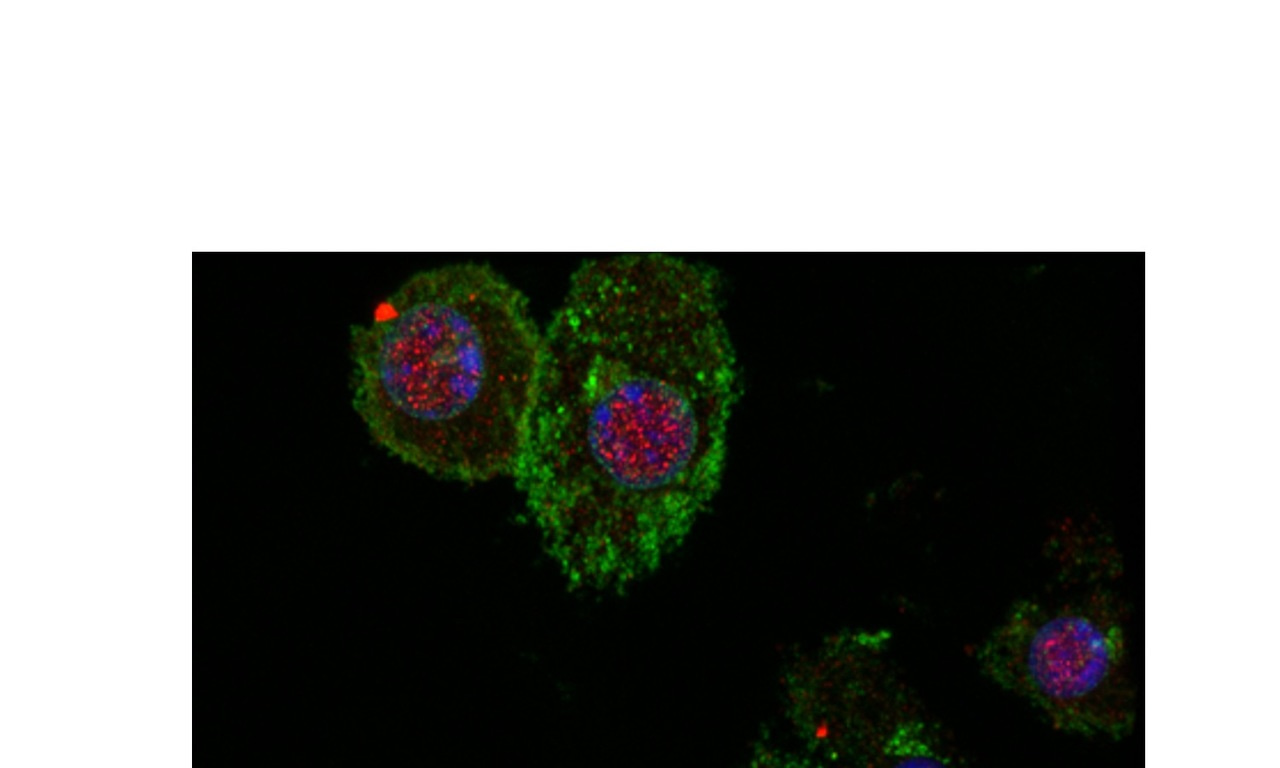
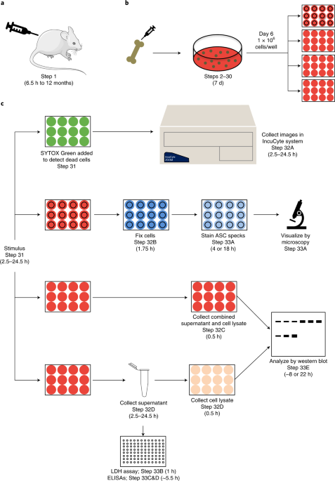
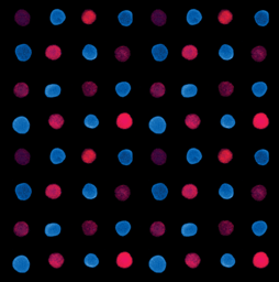
In our recent article in Nature Protocols, we describe methods that allow for the analysis of multiple inflammasome readouts from samples obtained in parallel using a single isolation of cells, i.e. from a single experiment (Figure). Our workflow assesses inflammasome activation via the formation of the ASC speck, cleavage of caspase-1 and gasdermin D, and release of IL-1β, IL-18, and caspase-1, as well as monitoring cell death by measuring lactate dehydrogenase release from the cell and performing real-time analysis by imaging. Additionally, our protocol describes a number of disease models and stimulating conditions that are associated with inflammasome activation both in vivo and in vitro and explains how to successfully use these models. These include bacterial, viral, and fungal infection, inflammatory disease models, and cancer models.
The most exciting aspect of our protocol is its versatility. In addition to all the disease models we describe, the inflammasome readouts can be coupled with any experimental trigger or disease model of interest to determine whether it induces activation. The readouts can also be expanded to answer other molecular questions and assess other forms of cell death, which will also be detected in the assays. For example, we have recently found extensive crosstalk between pyroptosis, apoptosis, and necroptosis, leading us to characterize PANoptosis as a unique cell death regulated by the PANoptosome, which provides a molecular scaffold that allows for interactions and activation of the machinery required for inflammasome/pyroptosis, apoptosis, and necroptosis. Since inflammasome components are core members of the PANoptosome, these methods can also be extended to evaluate PANoptosis.
Using our method provides insights into the role of inflammasome activation and cell death in any number of disease processes. This is critical for identifying new therapeutic targets and developing strategies to control the appropriate activation of the inflammasome while inhibiting aberrant activation. We hope that our protocol will allow researchers to make important progress to improve our ability to treat infectious, inflammatory, and metabolic diseases and cancers.
Follow the Topic
-
Nature Protocols

This journal publishes secondary research articles and covers new techniques and technologies, as well as established methods, used in all fields of the biological, chemical and clinical sciences.

Please sign in or register for FREE
If you are a registered user on Research Communities by Springer Nature, please sign in