JMCR: Clinical Reasoning from Case Reports
Published in Neuroscience and Biomedical Research
Explore the Research
Rare isolated basal vein of Rosenthal thrombosis causing thalamic haemorrhage during intensive-phase antituberculous therapy: a case report
Based on: Walikonthage BN, Udurawana AMJB, Kumarihamy P, et al. Rare isolated basal vein of Rosenthal thrombosis causing thalamic haemorrhage during intensive-phase antituberculous therapy: a case report. Journal of Medical Case Reports. 2026.
DOI: 10.1186/s13256-026-06528-4.
The Case
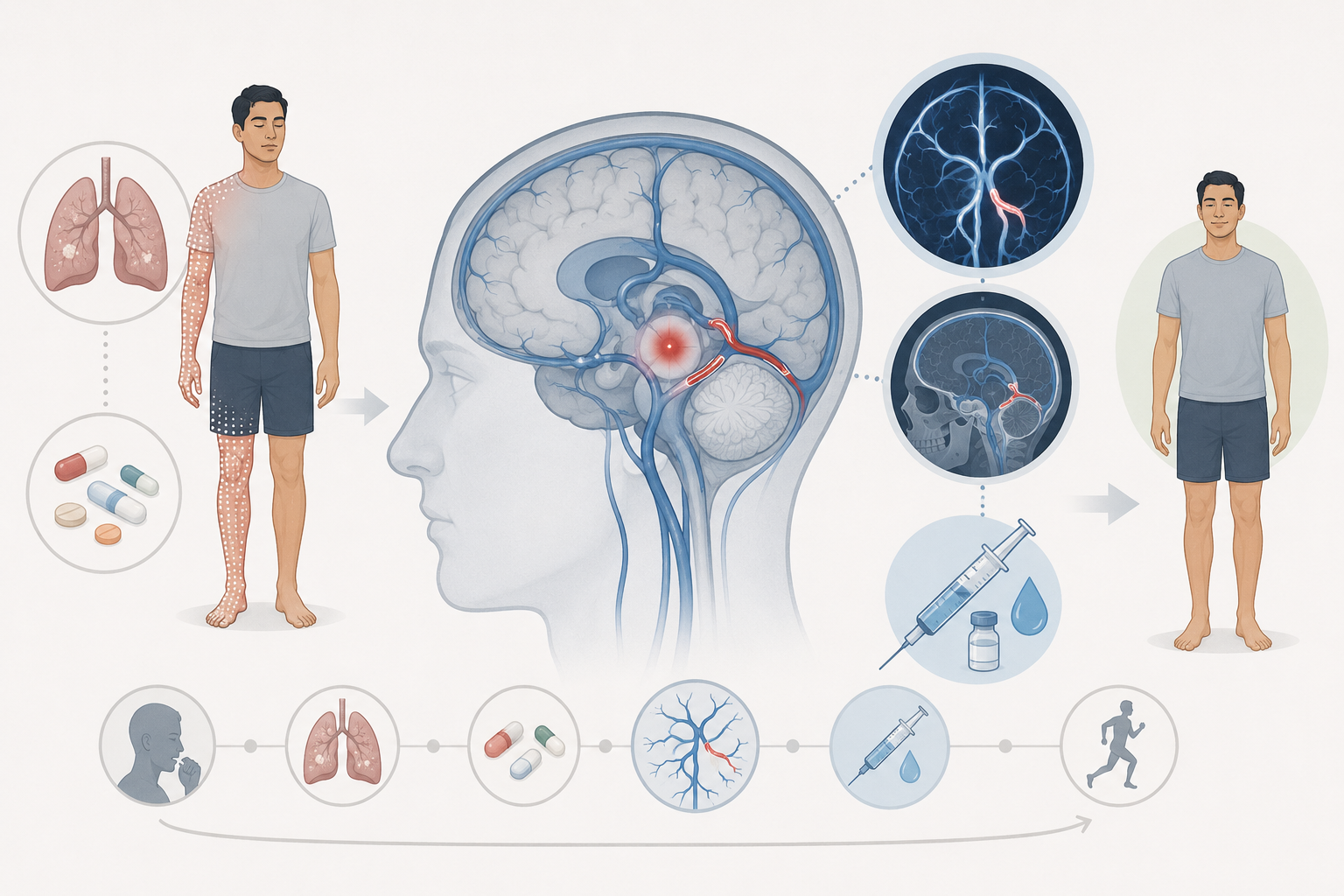
A 23-year-old Sri Lankan man with recently diagnosed extrapulmonary pleural tuberculosis presented with sudden right-sided numbness involving the upper and lower extremities. His symptoms had been present for approximately one hour and were purely sensory, without weakness.
Two weeks earlier, he had started intensive-phase antituberculous therapy with isoniazid, rifampicin, pyrazinamide, and ethambutol.
He had no hypertension, smoking history, alcohol use, known coagulopathy, prior thromboembolic disease, or family history of bleeding or thrombosis.
On examination, he was alert, afebrile, and hemodynamically stable. Blood pressure was 126/76 mmHg. Neurologic examination showed isolated right-sided sensory impairment, more prominent in the upper limb, with preserved strength, reflexes, joint position sense, and vibration. There were no cranial nerve abnormalities, meningeal signs, or evidence of raised intracranial pressure.
His NIH Stroke Scale score was 1.
Clinical Reasoning Pause 1
A young patient has an acute hemisensory syndrome. Where is the lesion?
The combination of right face- and body-independent hemisensory symptoms without weakness strongly localizes to a left-sided central sensory pathway.
A lesion involving the left thalamus is particularly important to consider.
At this stage, the differential diagnosis includes:
-
small thalamic ischemic stroke,
-
thalamic hemorrhage,
-
vascular malformation,
-
cerebral venous thrombosis with venous infarction or hemorrhage,
-
inflammatory or infectious vascular disease,
-
coagulopathy,
-
and less commonly a structural lesion.
Because the patient is only 23 years old and has no history of hypertension, a conventional hypertensive deep intracerebral hemorrhage would be unusual.
First Imaging Study
Noncontrast CT demonstrated an isolated left thalamic intracerebral hemorrhage with minimal surrounding edema, without intraventricular extension or midline shift.
The CT image presented in Figure 1 of the report shows the small focal hemorrhage within the left thalamus.
Clinical Reasoning Pause 2
The CT shows a thalamic hemorrhage. Is the diagnosis complete?
No.
The critical reasoning step is to distinguish:
Where is the hemorrhage?
from:
Why did the hemorrhage occur?
In an older patient with longstanding hypertension, a thalamic hemorrhage might reasonably suggest hypertensive small-vessel disease.
In this 23-year-old patient, however:
-
blood pressure was normal,
-
coagulation studies were normal,
-
there was no known bleeding disorder,
-
and there were no conventional vascular risk factors.
The hemorrhage therefore requires an etiologic investigation.
Particularly important possibilities include:
-
arteriovenous malformation or another arterial vascular lesion,
-
cerebral venous thrombosis,
-
thrombophilia,
-
vasculitis,
-
infectious or inflammatory vascular disease.
The recent diagnosis of active tuberculosis adds an important clue: tuberculosis may be associated with a systemic prothrombotic state.
Initial Laboratory Evaluation
The patient's:
-
complete blood count,
-
platelet count,
-
PT,
-
aPTT,
-
INR,
-
CRP,
-
ESR,
-
renal function,
-
liver function,
-
and serum electrolytes
were within normal limits.
Further evaluation included antiphospholipid antibodies, ANA, protein C, protein S, homocysteine, HIV, hepatitis B and C testing, and peripheral blood film. These studies were unrevealing.
Factor V Leiden mutation, prothrombin gene mutation, and antithrombin III levels could not be tested because of resource limitations.
Arterial Imaging
CT angiography showed no aneurysm or vascular malformation.
At this point, an arterial explanation for the hemorrhage became less likely.
Contrast-enhanced CT subsequently demonstrated nonvisualization of the left basal vein of Rosenthal.
That finding shifted the diagnostic reasoning toward the cerebral venous circulation.
Clinical Reasoning Pause 3
Why does the basal vein of Rosenthal matter?
The basal vein of Rosenthal is an important component of the deep cerebral venous system. It drains structures that include the:
-
thalamus,
-
basal ganglia,
-
medial temporal region,
before ultimately draining toward the vein of Galen.
Thrombosis of this vein can therefore produce deep cerebral parenchymal abnormalities, including hemorrhagic lesions.
Deep cerebral venous thrombosis more commonly involves the internal cerebral veins, vein of Galen, or straight sinus. Isolated basal vein of Rosenthal thrombosis is exceptionally uncommon.
Because routine arterial imaging may not adequately demonstrate the venous abnormality, dedicated venographic imaging can be decisive.
The Diagnostic Study
CT venography demonstrated:
-
normal visualization of the right basal vein of Rosenthal,
-
nonvisualization of the left basal vein of Rosenthal,
consistent with isolated thrombosis of the left basal vein of Rosenthal.
The venographic images in Figures 2 and 3 demonstrate this asymmetry, with preservation of the contralateral basal vein and absence of the expected left-sided venous structure.
Final Diagnosis
Isolated left basal vein of Rosenthal thrombosis causing a left thalamic hemorrhage in a patient undergoing intensive-phase treatment for pleural tuberculosis.
Clinical Reasoning Pause 4
How can a venous thrombosis produce an intracerebral hemorrhage?
Cerebral venous thrombosis impairs venous outflow.
The resulting sequence may include:
venous obstruction → increased venous and capillary pressure → impaired cerebral perfusion → disruption of the blood–brain barrier → edema → venous infarction → hemorrhagic transformation.
Thus, hemorrhage in cerebral venous thrombosis is mechanistically different from primary arterial rupture.
This distinction matters because the treatment strategy is also different.
Why Did This Patient Develop Thrombosis?
The authors considered tuberculosis-associated hypercoagulability the most plausible unifying mechanism.
Tuberculosis may promote thrombosis through several proposed mechanisms described in the report:
-
systemic inflammation,
-
endothelial dysfunction,
-
cytokine-mediated increases in procoagulant activity,
-
increased fibrinogen and other clotting factors,
-
altered natural anticoagulant pathways,
-
platelet activation,
-
and reactive changes within the coagulation system.
The authors also discuss two possible additional contributors.
Early treatment-related inflammatory response
The thrombosis occurred after approximately 14 days of intensive antituberculous therapy. The report raises the possibility that an early inflammatory response associated with treatment, including a possible tuberculosis-associated immune reconstitution inflammatory syndrome, could have contributed.
However, the authors appropriately describe this as a possible contributing mechanism rather than a definitive explanation.
Rifampicin
The report also notes that rifampicin-containing regimens have been associated with thromboembolic events in some studies and case reports.
Again, the case itself cannot establish that rifampicin caused the thrombosis.
The strongest source-supported conclusion is therefore that active tuberculosis-associated hypercoagulability was considered the most plausible underlying predisposition, with treatment-related inflammatory mechanisms or rifampicin as possible additional contributors.
Clinical Reasoning Pause 5
There is intracranial hemorrhage. Should the patient receive anticoagulation?
This is the counterintuitive but essential management principle in cerebral venous thrombosis.
The hemorrhage is occurring because of venous obstruction and congestion, not because anticoagulation has caused spontaneous arterial bleeding.
For that reason, the authors emphasize that anticoagulation remains the cornerstone of cerebral venous thrombosis treatment even when intracerebral hemorrhage is present.
Management
Diagnosis was delayed because cerebral venous thrombosis was not initially suspected and because of resource limitations. CT venography was ultimately performed on hospital day 10.
Once the diagnosis was established:
-
therapeutic subcutaneous enoxaparin was started,
-
antituberculous treatment was continued uninterrupted,
-
serial neurologic examinations were performed.
Repeat imaging approximately 3 weeks later demonstrated no progression of the hemorrhage.
At 3 weeks, bridging anticoagulation with enoxaparin and warfarin was initiated.
The patient was discharged on hospital day 31 taking warfarin 10 mg daily with a target INR of 2–3.
Six weeks after discharge, anticoagulation was changed to rivaroxaban 20 mg daily.
Outcome
Neurologic symptoms gradually improved during hospitalization.
At 3-month follow-up, the patient had marked neurologic improvement and had experienced no recurrent thrombotic events.
Follow-up contrast-enhanced CT and venography could not be obtained because of resource limitations.
What Made This Case Difficult?
1. The initial image looked like a primary hemorrhage
The first CT demonstrated a thalamic hemorrhage. Without consideration of the underlying cause, the diagnostic process could have ended there.
2. The patient had very mild neurologic findings
His NIHSS score was only 1, and his deficit was purely sensory.
3. Routine laboratory studies were normal
Normal PT, aPTT, INR, CRP, and ESR did not exclude a prothrombotic disorder.
4. The thrombosis involved an exceptionally unusual vein
Deep cerebral venous thrombosis itself is uncommon, while isolated basal vein of Rosenthal thrombosis is particularly rare.
5. Dedicated venous imaging was required
CT angiography excluded arterial abnormalities but did not establish the diagnosis. CT venography provided the critical evidence.
Diagnostic Pitfalls
Pitfall 1: Equating hemorrhage location with hemorrhage etiology
A deep hemorrhage does not automatically mean hypertensive small-vessel disease, particularly in a young normotensive patient.
Pitfall 2: Stopping after a negative CT angiogram
CTA can exclude many arterial causes but does not substitute for dedicated venography when cerebral venous thrombosis remains possible.
Pitfall 3: Being reassured by normal coagulation tests
Routine coagulation studies do not exclude cerebral venous thrombosis or a systemic prothrombotic state.
Pitfall 4: Avoiding anticoagulation because blood is visible on CT
In cerebral venous thrombosis, hemorrhagic venous infarction does not by itself eliminate the indication for anticoagulation.
Pitfall 5: Attributing causation too strongly
This case establishes an association between active tuberculosis, antituberculous treatment, and venous thrombosis. It does not prove that tuberculosis, TB-IRIS, or rifampicin individually caused the thrombosis.
Clinical Pearls
Pearl 1: A spontaneous thalamic hemorrhage in a young normotensive patient warrants investigation for an underlying vascular cause.
Pearl 2: Deep cerebral venous thrombosis can mimic primary intracerebral hemorrhage.
Pearl 3: Isolated basal vein of Rosenthal thrombosis should be considered when a deep cerebral lesion does not have an adequate arterial explanation.
Pearl 4: Normal PT, aPTT, INR, inflammatory markers, and common thrombophilia studies do not rule out cerebral venous thrombosis.
Pearl 5: CT or MR venography may be necessary when venous thrombosis is suspected despite nondiagnostic arterial imaging.
Pearl 6: Active tuberculosis may create a systemic prothrombotic environment even without tuberculous meningitis.
Pearl 7: Anticoagulation remains central to treatment of cerebral venous thrombosis despite associated intracerebral hemorrhage.
CARE-Style Clinical Timeline
| Time | Clinical event |
|---|---|
| 2 weeks before presentation | Pleural tuberculosis diagnosed; isoniazid, rifampicin, pyrazinamide, and ethambutol started |
| Day 0 | Acute right-sided numbness; NIHSS 1 |
| Day 0 | Noncontrast CT: isolated left thalamic hemorrhage |
| Initial evaluation | Normal blood pressure, routine coagulation studies, inflammatory markers, and major acquired thrombophilia testing |
| Subsequent imaging | CT angiography: no arterial vascular lesion |
| Further CT imaging | Nonvisualization of left basal vein of Rosenthal suspected |
| Hospital day 10 | CT venography confirmed isolated left basal vein of Rosenthal thrombosis |
| Day 10 | Therapeutic enoxaparin initiated; tuberculosis therapy continued |
| Approximately 3 weeks | Repeat imaging showed no hemorrhagic progression; enoxaparin/warfarin bridging performed |
| Day 31 | Discharged on warfarin with target INR 2–3 |
| 6 weeks after discharge | Changed to rivaroxaban 20 mg daily |
| 3 months | Marked neurologic improvement; no recurrent thrombosis |
Test Your Clinical Reasoning
Question 1
A 23-year-old normotensive patient develops acute hemisensory loss, and CT demonstrates a small unilateral thalamic hemorrhage. CT angiography is normal. Which investigation is most important next if a venous cause is suspected?
A. Repeat noncontrast CT only
B. Carotid ultrasound
C. CT or MR venography
D. Electroencephalography
E. Lumbar puncture
Answer: C. CT or MR venography
Explanation: Dedicated venographic imaging is required to evaluate the deep cerebral venous system. In this case, CT venography demonstrated isolated thrombosis of the basal vein of Rosenthal.
Question 2
Which feature most strongly argues against simply labeling this patient's thalamic hemorrhage as a conventional hypertensive hemorrhage?
A. Male sex
B. Right-sided sensory symptoms
C. Age 23 years with normal blood pressure
D. NIHSS score of 1
E. Absence of headache
Answer: C. Age 23 years with normal blood pressure
Explanation: Deep hemorrhage may occur with hypertension, but a spontaneous thalamic hemorrhage in a young normotensive patient should prompt evaluation for alternative vascular and hematologic causes.
Question 3
Which mechanism best explains intracerebral hemorrhage associated with cerebral venous thrombosis?
A. Rupture of a penetrating artery
B. Venous congestion producing increased capillary pressure and hemorrhagic venous infarction
C. Thrombocytopenia caused by tuberculosis
D. Direct invasion of the cerebral arteries by rifampicin
E. Embolism from the carotid artery
Answer: B. Venous congestion producing increased capillary pressure and hemorrhagic venous infarction
Explanation: Venous obstruction raises venous and capillary pressure, disrupts the blood–brain barrier, and can cause venous infarction with hemorrhagic transformation.
Question 4
Which statement about the relationship between tuberculosis and this patient's thrombosis is best supported by the case report?
A. Rifampicin definitively caused the thrombosis.
B. TB-IRIS was proven to be responsible.
C. Tuberculosis-associated hypercoagulability was considered the most plausible underlying predisposition.
D. Tuberculous meningitis directly invaded the cerebral veins.
E. The patient had a confirmed inherited thrombophilia.
Answer: C. Tuberculosis-associated hypercoagulability was considered the most plausible underlying predisposition.
Explanation: The authors discuss systemic inflammation and hypercoagulability from active tuberculosis as the most plausible unifying explanation. TB-IRIS and rifampicin are discussed only as possible additional contributors.
Question 5
What is the key management principle demonstrated by this case?
A. Intracerebral hemorrhage is an absolute contraindication to anticoagulation.
B. Cerebral venous thrombosis associated with hemorrhage can still require therapeutic anticoagulation.
C. Antituberculous therapy should always be discontinued after thrombosis.
D. Surgery is first-line treatment for isolated basal vein thrombosis.
E. Anticoagulation should begin only after complete radiographic resolution of the hemorrhage.
Answer: B. Cerebral venous thrombosis associated with hemorrhage can still require therapeutic anticoagulation.
Explanation: The patient was treated with therapeutic anticoagulation despite the thalamic hemorrhage and improved without radiographic progression of bleeding.
Clinical Take-Home Message
When a young patient without hypertension or coagulopathy presents with a deep intracerebral hemorrhage, the diagnostic process should not stop at the hemorrhage itself.
Ask what caused it.
In this case, a seemingly straightforward thalamic hemorrhage was actually the consequence of isolated thrombosis of the basal vein of Rosenthal. The diagnosis required dedicated venous imaging, and identifying the venous mechanism fundamentally changed management.
The central lesson is:
In an atypical intracerebral hemorrhage—particularly in a young patient with a prothrombotic condition—look beyond the blood and examine the veins.
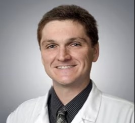
Richard Alan Rison is the Editor-in-Chief of Journal of Medical Case reports. He is also an associate neurology editor (editorial board) for BMC Neurology, and the former lead editor for case reports of BMC Research Notes (currently on the editorial board). His scholarly work focuses on medical case reporting, reporting standards, and editorial methodology. Dr. Rison participated in the development and dissemination of the CARE guidelines for clinical case reporting and has authored numerous publications addressing both neurological disorders and the role of case reports in advancing medical knowledge. Dr. Rison practices general neurology and served as the founding medical director of the PIH Health Hospital-Whittier Stroke Program and the PIH Health Hospital-Whittier Non-Invasive Vascular Laboratory, is a clinical assistant professor of neurology at the University of Southern California Keck School of Medicine and Los Angeles County Medical Center, and is a Fellow of the American Academy of Neurology, the American Neurological Association, and the American Association of Neuromuscular and Electrodiagnostic Medicine. Dr Rison is board-certified by the American Board of Psychiatry and Neurology in neurology and vascular neurology, and neurocritical care and neuroimaging by the United Council of Neurologic Subspecialties. He is also board-certified by the American Board of Electrodiagnostic Medicine in electrodiagnostic medicine. Dr. Rison is a former president of the Los Angeles Neurological Society.
Follow the Topic
-
Journal of Medical Case Reports
This journal will consider any original case report that expands the field of general medical knowledge, and original research relating to case reports.