JMCR: Clinical Reasoning from Case Reports
Published in Neuroscience and Biomedical Research
Explore the Research
Histopathological analysis of a retrieved cerebral thrombus revealing occult lung adenocarcinoma: a case report
A 76-year-old White European man with a history of tobacco use presented with the sudden onset of left-sided hemiplegia, hemianopia, and dysarthria. His NIHSS score was 18.
Non-contrast CT demonstrated early ischaemic changes with an ASPECTS of 7. CT angiography showed an occlusion of the right middle cerebral artery, while CT perfusion demonstrated a substantial penumbral region with a favourable mismatch profile. Intravenous thrombolysis was not given because of partially established infarction.
A pretreatment chest radiograph showed a left retrocardiac opacity, but its appearance was nonspecific and was not initially considered suggestive of malignancy. Angiography confirmed the cerebral arterial occlusion, and mechanical thrombectomy achieved partial reperfusion with a final mTICI score of 2b.
Clinical Reasoning Pause 1
What is the immediate diagnosis?
This is an acute right middle cerebral artery ischaemic stroke caused by a large-vessel occlusion.
At this point, however, the mechanism of the embolism remains unknown.
What should we be thinking about?
The immediate priority is reperfusion, but etiologic reasoning begins in parallel. In an older patient with an embolic-appearing large-vessel stroke, possible mechanisms include a cardiac embolic source, cancer-associated hypercoagulability, non-bacterial thrombotic endocarditis, or less commonly direct tumour embolisation.
There was nothing at presentation that clearly identified malignancy as the cause. Even the abnormal chest radiograph was nonspecific.
Key reasoning point: Do not allow an initially ordinary-appearing large-vessel stroke to prematurely determine the eventual mechanism.
The next clue
Two days after thrombectomy, brain MRI demonstrated an extensive acute infarction in the right middle cerebral artery territory without evidence of brain metastases.
Continuous telemetry and a subsequent 72-hour Holter study showed no atrial fibrillation or other clinically important arrhythmia. Transthoracic echocardiography demonstrated no valvular vegetation, intracardiac thrombus, or other major cardioembolic source.
Laboratory studies were largely unrevealing apart from a D-dimer concentration of 1,476 ng/mL, only moderately above the reference range of 0–500 ng/mL.
Clinical Reasoning Pause 2
Does the moderately elevated D-dimer make cancer-associated stroke unlikely?
Not necessarily.
Cancer-associated stroke is often associated with pronounced D-dimer elevation, particularly when the mechanism is systemic hypercoagulability. But cancer-associated stroke is not a single pathophysiologic entity.
The authors distinguish several mechanisms, including:
-
cancer-associated hypercoagulability,
-
non-bacterial thrombotic endocarditis,
-
direct vascular or tumour involvement,
-
and treatment-related vascular complications.
Direct tumour embolism is particularly unusual and is thought to account for less than 1% of cancer-associated strokes.
A modest D-dimer elevation therefore cannot exclude a malignant mechanism, especially if the embolic material itself is tumour rather than predominantly fibrin-rich thrombus.
The diagnostic turning point
The thrombectomy specimen was sent for histopathological examination.
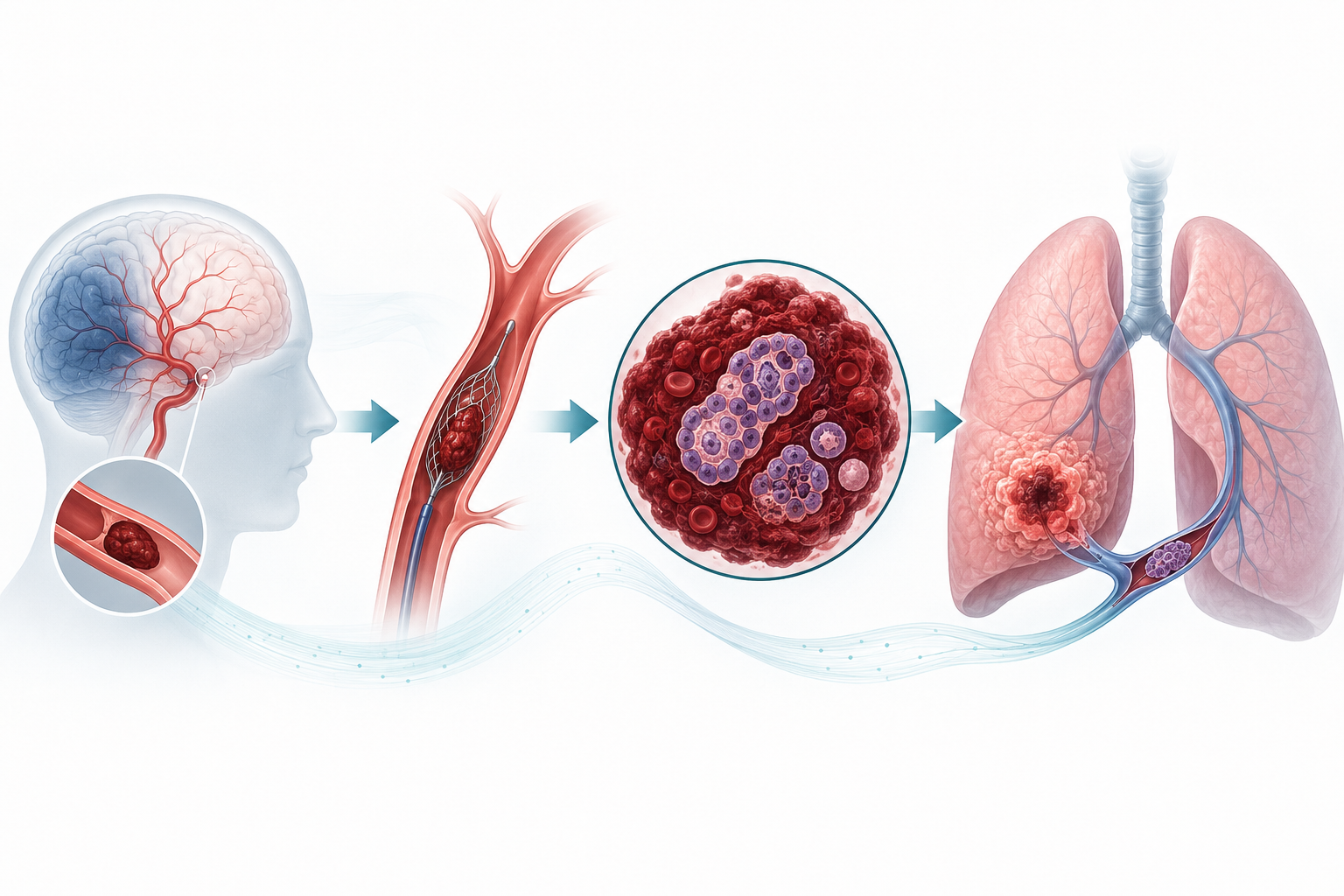
Instead of showing only conventional thrombotic material, the specimen contained nests of malignant epithelial cells consistent with non-small-cell carcinoma.
Immunohistochemical analysis demonstrated:
-
CK7 positivity,
-
weak, patchy TTF-1 expression,
-
negative P40 staining,
-
negative synaptophysin staining.
These findings supported poorly differentiated pulmonary adenocarcinoma.
PD-L1 expression was high, with a tumour proportion score of 90%. Next-generation sequencing detected a pathogenic TP53 exon 5 mutation but no potentially actionable alterations in the tested EGFR, BRAF, ALK, ROS1, KRAS, RET, MET, ERBB2, or NTRK genes.
Clinical Reasoning Pause 3
What does finding malignant epithelial cells inside the retrieved cerebral embolus mean?
This changes the problem fundamentally.
The stroke is no longer simply an embolic stroke occurring in a patient who happens to have cancer. The thrombectomy specimen itself provides direct evidence that malignant tissue participated in the cerebral embolic event.
The next question becomes:
Where did the tumour embolus originate, and how did it enter the systemic arterial circulation?
Finding the source
Thoracic CT demonstrated a 9 × 7 × 8 cm heterogeneous left lower-lobe mass with central necrosis, mediastinal lymphadenopathy, and, crucially, invasion of a pulmonary vein.
The malignancy was staged as cT4N2M1c, stage IV poorly differentiated pulmonary adenocarcinoma.
Pulmonary vein invasion provides an anatomically plausible pathway by which tumour material can enter the left-sided circulation and embolise directly to a cerebral artery.
The combination of:
-
malignant epithelial cells within the cerebral thrombectomy specimen,
-
a pulmonary adenocarcinoma invading a pulmonary vein,
-
absence of atrial fibrillation during the documented monitoring period,
-
and no major embolic source detected by transthoracic echocardiography
strongly supported direct cerebral tumour embolism from the lung malignancy.
But is the mechanism completely proven?
Not entirely.
Non-bacterial thrombotic endocarditis remains an important alternative or potentially concomitant mechanism of cancer-associated stroke.
Transthoracic echocardiography showed no vegetations, but transoesophageal echocardiography was not performed. The authors therefore appropriately acknowledge that occult NBTE could not be definitively excluded.
This is an important distinction in clinical reasoning:
Strong evidence for one mechanism does not justify claiming exclusion of another when the necessary diagnostic test was not performed.
Management and outcome
Because no actionable driver alteration was identified and PD-L1 expression was high, the patient began treatment with pembrolizumab.
Neurologically, he initially improved. At discharge his NIHSS score had decreased from 18 to 7, and his modified Rankin Scale score was reported as 2.
Thirty days later, however, he was readmitted with fever, behavioural changes, left-sided sensory impairment, and hyperphagia. Brain imaging now demonstrated frontal metastases.
His neurological and systemic condition deteriorated rapidly, and he died 51 days after the index stroke.
Reconstructing the diagnostic pathway
Presentation
Sudden left hemiplegia, hemianopia, and dysarthria
NIHSS 18
↓
Neurovascular imaging
Right middle cerebral artery occlusion
ASPECTS 7 with favourable perfusion mismatch
↓
Acute treatment
Mechanical thrombectomy
mTICI 2b
↓
Initial etiologic evaluation
No atrial fibrillation on telemetry or 72-hour Holter
No major embolic source on transthoracic echocardiography
D-dimer only moderately elevated
↓
Unexpected finding
Malignant epithelial cells within the retrieved cerebral thrombus
↓
Pathological characterization
CK7 positive
Weak patchy TTF-1 positive
P40 negative
Synaptophysin negative
↓
Search for primary tumour
Large left lower-lobe lung mass with pulmonary vein invasion
↓
Final clinicopathological interpretation
Poorly differentiated pulmonary adenocarcinoma with direct cerebral tumour embolisation strongly supported
Why this case matters
Most cancer-associated strokes are attributed to hypercoagulability rather than macroscopic tumour embolisation. Direct tumour embolism is considered exceptionally uncommon.
The diagnostic lesson in this case is that the material removed during thrombectomy was not merely therapeutic debris—it became a tissue diagnosis.
In the authors' institution, retrieved thrombectomy specimens are routinely submitted for histopathological evaluation. They appropriately stop short of recommending universal pathological examination of every thrombectomy specimen, but suggest that histopathology may provide additional diagnostic information in selected patients with embolic stroke of undetermined source, atypical features, or otherwise unexplained embolism.
The case also illustrates why D-dimer should be interpreted in the context of mechanism. Marked elevation may suggest hypercoagulability-mediated cancer-associated stroke, but a tumour fragment embolising directly from a pulmonary vein does not necessarily produce the same laboratory phenotype.
Clinical Pearls
1. Cancer-associated stroke is mechanistically heterogeneous.
Hypercoagulability is common, but NBTE and direct tumour embolisation are distinct possibilities.
2. The thrombectomy specimen can contain diagnostic information beyond clot composition.
Malignant cells within retrieved embolic material may reveal an unsuspected malignancy.
3. A modest D-dimer level does not exclude malignancy-associated stroke.
The relevance of D-dimer depends partly on the underlying embolic mechanism.
4. Pulmonary vein invasion provides a direct route for systemic tumour embolisation.
A lung tumour invading a pulmonary vein can potentially release malignant tissue directly into the systemic arterial circulation.
5. Negative transthoracic echocardiography does not completely exclude NBTE.
In this case, TEE was not performed, so occult NBTE remained a limitation of the etiologic assessment.
6. Stroke may precede recognition of the malignancy.
The cerebral embolic event was the clinical event that ultimately led to identification of the patient's advanced pulmonary adenocarcinoma.
Pitfalls to Avoid
Pitfall 1: Assuming every cancer-associated stroke is caused by hypercoagulability
Cancer-associated stroke describes an association, not a single mechanism.
Pitfall 2: Using D-dimer as a rule-out test for tumour embolism
The patient's D-dimer was elevated only moderately despite compelling pathological evidence of malignant embolic material.
Pitfall 3: Ignoring the retrieved thrombectomy material
Pathological examination may occasionally transform an apparently cryptogenic embolic stroke into a specific clinicopathological diagnosis.
Pitfall 4: Declaring NBTE excluded after a negative TTE
The authors specifically acknowledge that the absence of TEE prevented complete exclusion of occult NBTE.
Pitfall 5: Confusing tumour embolism with cerebral metastasis
The MRI obtained two days after thrombectomy showed acute infarction without brain metastases. Metastases appeared later during the clinical course.
CARE-Style Timeline
| Time | Clinical event |
|---|---|
| Day 0 | Acute left hemiplegia, hemianopia, and dysarthria; NIHSS 18 |
| Day 0 | CT/CTA/CT perfusion demonstrate right MCA occlusion, ASPECTS 7, and favourable mismatch |
| Day 0 | Mechanical thrombectomy performed; final mTICI 2b |
| Day 2 | MRI confirms extensive right MCA-territory acute infarction without metastases |
| Early evaluation | Telemetry and 72-hour Holter show no atrial fibrillation; TTE reveals no major cardioembolic source |
| After thrombectomy | Histopathology identifies malignant epithelial cells in retrieved embolic material |
| Day 5 | Thoracic CT demonstrates 9 × 7 × 8 cm left lower-lobe mass with pulmonary vein invasion |
| Oncological workup | Poorly differentiated pulmonary adenocarcinoma staged cT4N2M1c; PD-L1 tumour proportion score 90% |
| Treatment | Pembrolizumab initiated |
| Discharge | Neurological improvement to NIHSS 7; mRS 2 |
| Approximately Day 30 | Readmission with new neurological/behavioural symptoms; frontal brain metastases identified |
| Day 51 | Death following rapid neurological and systemic deterioration |
Test Your Clinical Reasoning
Question 1
A 76-year-old man undergoes thrombectomy for an acute MCA occlusion. Which subsequent finding most directly supports tumour embolism rather than conventional cancer-associated hypercoagulability?
A. D-dimer of 1,476 ng/mL
B. History of tobacco use
C. Malignant epithelial cells within the retrieved embolus
D. Large MCA-territory infarction
E. Absence of atrial fibrillation
Answer: C. Malignant epithelial cells within the retrieved embolus
Explanation: Demonstration of malignant cells within the cerebral embolic material provides direct pathological evidence that tumour tissue participated in the embolic event. The other findings may contribute to etiologic reasoning but are not specific for tumour embolisation.
Question 2
Which finding provides the most plausible anatomical route for tumour cells from this patient's lung malignancy to reach the cerebral circulation?
A. Mediastinal lymphadenopathy
B. Central tumour necrosis
C. Pulmonary vein invasion
D. High PD-L1 expression
E. TP53 mutation
Answer: C. Pulmonary vein invasion
Explanation: Invasion of a pulmonary vein provides direct access to the left-sided systemic circulation, permitting tumour material to embolise into systemic arteries, including the cerebral circulation.
Question 3
What is the most appropriate interpretation of the patient's moderately elevated D-dimer?
A. It excludes cancer-associated stroke
B. It proves NBTE
C. It proves systemic cancer-associated hypercoagulability
D. It does not exclude direct tumour embolisation
E. It excludes advanced malignancy
Answer: D. It does not exclude direct tumour embolisation
Explanation: Markedly elevated D-dimer concentrations are more characteristic of hypercoagulability-mediated cancer-associated stroke. Direct embolisation of tumour material may occur without marked D-dimer elevation.
Question 4
Which limitation prevents complete exclusion of non-bacterial thrombotic endocarditis in this patient?
A. MRI was obtained after thrombectomy
B. Only 72 hours of Holter monitoring were performed
C. Transoesophageal echocardiography was not performed
D. PD-L1 expression was elevated
E. The cerebral artery was only partially recanalised
Answer: C. Transoesophageal echocardiography was not performed
Explanation: TTE demonstrated no vegetation, but the authors emphasize that without TEE, occult NBTE could not be completely excluded.
Question 5
What was the most important diagnostic contribution of histopathological examination of the thrombectomy specimen in this case?
A. It determined the mTICI score
B. It established the extent of cerebral infarction
C. It provided the first major clue to an occult pulmonary malignancy
D. It excluded cerebral metastases
E. It demonstrated atrial fibrillation as the embolic mechanism
Answer: C. It provided the first major clue to an occult pulmonary malignancy
Explanation: Identification of malignant epithelial cells in the retrieved material prompted investigation for an underlying primary malignancy, ultimately revealing advanced pulmonary adenocarcinoma with pulmonary vein invasion.
Clinical Take-Home Message
When an embolic stroke remains unexplained, the retrieved thrombus itself may contain the answer. This case demonstrates how malignant cells identified during pathological examination of a thrombectomy specimen led to recognition of an occult lung adenocarcinoma invading a pulmonary vein and strongly supported direct cerebral tumour embolisation. A modest D-dimer elevation should not exclude this mechanism, and competing mechanisms such as NBTE should be excluded as completely as the available investigation permits.
Source
Larrañaga C, Albajar I, Juaristi-Abaunz M, Marta-Enguita J. Histopathological analysis of a retrieved cerebral thrombus revealing occult lung adenocarcinoma: a case report. Journal of Medical Case Reports. 2026. doi:10.1186/s13256-026-06487-w.
Journal of Medical Case Reports is the world's first international, PubMed-listed medical journal devoted to publishing case reports from all medical disciplines and will consider any original case report that expands the field of general medical knowledge, and original research relating to case reports. The journal is open access and strongly endorses the CARE guidelines for case reports, requiring authors to submit populated CARE checklists with submissions to improve transparency in reporting.

Richard Alan Rison is the interim Editor-in-Chief of Journal of Medical Case reports. He is also an associate neurology editor (editorial board) for BMC Neurology, and the former lead editor for case reports of BMC Research Notes (currently on the editorial board). His scholarly work focuses on medical case reporting, reporting standards, and editorial methodology. Dr. Rison participated in the development and dissemination of the CARE guidelines for clinical case reporting and has authored numerous publications addressing both neurological disorders and the role of case reports in advancing medical knowledge. Dr. Rison practices general neurology and served as the founding medical director of the PIH Health Hospital-Whittier Stroke Program and the PIH Health Hospital-Whittier Non-Invasive Vascular Laboratory, is a clinical assistant professor of neurology at the University of Southern California Keck School of Medicine and Los Angeles County Medical Center, and is a Fellow of the American Academy of Neurology, the American Neurological Association, and the American Association of Neuromuscular and Electrodiagnostic Medicine. Dr Rison is board-certified by the American Board of Psychiatry and Neurology in neurology and vascular neurology, and neurocritical care and neuroimaging by the United Council of Neurologic Subspecialties. He is also board-certified by the American Board of Electrodiagnostic Medicine in electrodiagnostic medicine. Dr. Rison is a former president of the Los Angeles Neurological Society.
Follow the Topic
-
Journal of Medical Case Reports
This journal will consider any original case report that expands the field of general medical knowledge, and original research relating to case reports.