Living human brain slice cultures, Alzheimer’s disease and the power of philanthropy
Published in Neuroscience
Generating living human brain slice cultures
It’s 2019, and my optimistic “to do” list at the start of my fellowship has the following:
1) Learn how to make living human brain slices.
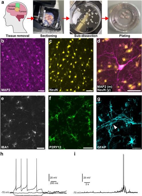
I was fortunate to visit my collaborator Dr Henner Koch, a pioneer of this method working in Germany. We scrubbed-in to a busy operating theatre and watched as the neurosurgeons carefully worked to remove a brain tumour from a patient covered in sterile drapes. They cut away a small piece of overlying brain to access the tumour below and, instead of placing this in the bin along with the other surgical debris, dropped it into our waiting collection bottle. The clock started. We hurried to the lab. The intense analysis of the sample began. Like working out how to cut a diamond out of a rock, the piece of brain must be perfectly orientated to preserve its unique layering. The brain is then sliced, at glacial speed, into 300 µm thick sections, plated onto culture membranes, then placed in an incubator. Live human brain in a dish. The opportunities are endless- but this is the first step in a long road.

Getting started in Edinburgh
In September 2019, I was at the start of my Race Against Dementia fellowship. This scheme, the brain-child of Formula One champion Sir Jackie Stewart, provides early career researchers with 5 years of funding to support “out of the box” projects working on dementia. Whilst my proposed project mostly relied on traditional laboratory methods, I had included a “long-shot” aim to establish a living human brain slice culture model of Alzheimer’s disease.
With the backing of the neurosurgical team, led by Prof. Paul Brennan, at the University of Edinburgh, we obtained all the necessary ethical approvals and started work. We then, immediately, went into months of COVID-19 related lockdowns. It wasn’t until early 2021 that myself, and my newly recruited PhD student Robert McGeachan, were able to regain access to the hospital to collect brain samples. Over many months, we painstakingly optimised collection procedures, cutting process and culture conditions. Through hours of conversations with Paul, our collaborator Dr Sam Booker, and other groups worldwide, we refined our pipeline to reliably generate, maintain and experiment on living human brain slice cultures.
Our proof of concept had worked, but we hit a serious bottle neck. Without clinical staff to perform the consent paperwork, we could only access a tiny fraction of the surgeries that were taking place. Our team of two was insufficient to maintain the cultures and sharing lab space with multiple groups meant we often struggled with contamination. We needed to adapt and expand.

Left: Dr Soraya Meftah processing live human brain using a vibratome. Right: Dr Robert McGeachan plating human brain slices into culture dishes.
A dedicated human slice culture laboratory
In July 2021, myself and Sir Jackie Stewart visited Sir James Dyson at his Malmesbury campus. His donation to Race Against Dementia had funded my fellowship and, now COVID-19 restrictions were easing, I was able to discuss my work with him, and his team of engineers, in person. I presented pilot data from our first human brain slice experiments, and discussed my plans for the future. At the end of the meeting, I was tasked with generating a proposal outlining how additional funding could accelerate our research.
What followed was a transformative £1 million pound donation from the James Dyson Foundation to establish the Dyson-RAD Dementia Research Acceleration Project. This enabled us to “buy time” in the EMERGE research nurse team at the hospital and provided funds for a postdoc (Dr Soraya Meftah) and PhD student (Lewis Taylor). Crucially, it enabled us to create our own dedicated human brain slice culture laboratory, filled with state-of-the-art equipment. The impact was immediate and lasting. Instead of obtaining a case every 2-3 months, we now receive brain tissue every week. Our full-strength team can process human brain tissue in an efficient and effective way. Specialised incubators provide optimal slice culture conditions and new equipment, such as electrophysiology rigs and multi-electrode arrays, enable us to perform more insightful experiments. Fully equipped, we could now focus on generating experimental data relating to Alzheimer’s disease. We are proud to share these results, now out in the journal Nature Communications.

Dr Claire Durrant, Sir James Dyson and Sir Jackie Stewart celebrating the launch of the Dyson-RAD Dementia Research Acceleration Project
Using sample diversity to our advantage
Unlike most experiments we conduct in the lab, where variables are eliminated wherever possible, working with human brain slices means you have to relinquish control. Every sample we receive is unique. They come from adults of all ages, different sexes, varied genetic background, and (depending on where the tumour is located) different regions of the brain. Using this variability to our advantage, we explored how the release of proteins related to Alzheimer’s disease (Amyloid beta (Aβ) and tau) are influenced by patient characteristics. Interestingly, we found the levels of Aβ released were impacted by the age of the patient, whilst tau release was highest in samples from a brain region called the temporal lobe. This brain region is affected early in Alzheimer’s disease, so understanding differences in tau release could help us uncover why this region is more vulnerable to damage.
Exploring the effect of Aβ at the synapse
In Alzheimer’s disease, it is known that loss of synapses, the connections between nerve cells that allow them to communicate, is the best correlate of cognitive decline. Too much Aβ is thought to be toxic to synapses, but evidence of this has been restricted to postmortem snapshots, animal models or cells. We wanted to see how changing Aβ levels impacts synapses in living human brain tissue. By using two different drugs, we were able to increase or decrease the natural production of Aβ. Interestingly, changes to Aβ concentration in either direction resulted in a loss of synapses- suggesting the brain requires a finely tuned sweet-spot of Aβ to keep synapses healthy. Excitingly, when we raised the concentration of Aβ, we found that the brain slices produced messenger molecules (RNA) to instruct the creation of new synaptic proteins, suggesting an attempt at repair.
In another experiment, we extracted “toxic” Aβ from the donated brains of people who had died with Alzheimer’s disease. We applied this extract directly to healthy brain slices and found, using a high-resolution imaging technique, that this Aβ binds to, and destroys synapses. Most interestingly, unlike when “normal” Aβ is increased, we did not see any attempts at repair at the RNA level. Understanding why living human brain responds differently to normal vs toxic Aβ may help us identify new drug targets to protect synapses in Alzheimer’s disease.
A future model for studying early stages of Alzheimer’s disease?
Without action, 1/3 of us will, unfortunately, die with dementia . As Alzheimer's disease takes decades to develop, many individuals will have pathology in their brain without clinical symptoms of the disease. By screening for Aβ plaques and tau tangles in the brain slices, we found evidence of early pathology in a subset of samples. Whilst the presence of these structures in a tiny brain sample does not guarantee that someone has, or would ever develop, Alzheimer’s disease, we are able to use these slices to explore the impact of plaques and tangles on brain tissue function. In the future we hope to use samples with pathology to test experimental treatments.
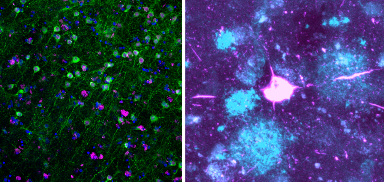
 and Aβ plaques (blue) in a live human brain slice")
Left: Living human brain slice culture after 7 days in culture. Neurons in green. Right: Tau tangle (pink) and Aβ plaques (blue) in a live human brain slice
A look to the future
The University of Edinburgh is now one of the leading centres in the world for using live human brain tissue for research. This is down to the hard work of an incredible team of scientists, research nurses, neurosurgeons and support staff united by the belief that the best model to study human brain diseases is the living human brain. The scale at which we can use this tissue is only possible because of the power of philanthropy. In a world where scientific funding is becoming more and more unstable, we are thankful to those who see the power of research, and act to help. We hope that our work will accelerate progress towards a world free from the heartbreak of dementia.

Members of the Edinburgh Living Human Brain Research Group include research nurses, scientists and neurosurgeons.
Follow the Topic
-
Nature Communications
An open access, multidisciplinary journal dedicated to publishing high-quality research in all areas of the biological, health, physical, chemical and Earth sciences.

Your space to connect: The Psychedelics Hub
A new Communities’ space to connect, collaborate, and explore research on Psychotherapy, Clinical Psychology, and Neuroscience!
Continue reading announcementRelated Collections
With Collections, you can get published faster and increase your visibility.
Women's Health
Publishing Model: Hybrid
Deadline: Ongoing
Biosensing
Publishing Model: Hybrid
Deadline: Jun 30, 2026

Please sign in or register for FREE
If you are a registered user on Research Communities by Springer Nature, please sign in
What a fascinating read! Thank you for sharing.