Myriad Mapping (MM) from atomic to intergalactic scales

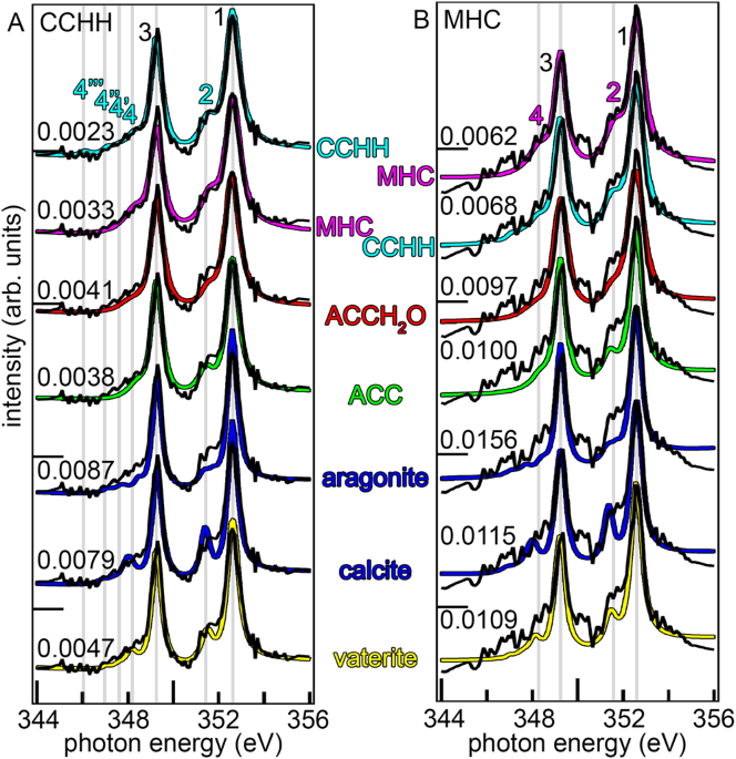
In a recent publication in Nature Communications (1), we discovered that the crystalline biominerals formed by corals and mollusks don’t only have liquid (2) and amorphous solid precursors (3-8), there are also crystalline precursors, albeit metastable, short-lives ones. The final mature biomineral in diverse coral skeletons and abalone nacre is crystalline, stable aragonite (CaCO3). But on the surface of forming nacre and coral skeletons we observed 2 amorphous and 2 crystalline metastable mineral phases, which, after approximately 1 day and a series of phase transitions, crystallize to aragonite (1).
The presence of 4 precursors and 1 mature phase in fresh, forming biominerals meant that the conventional RGB (9) or CMYK (9) imaging or mapping could not be used. A new method to quantitatively display all 5 phases had to be invented. Thus, we developed Myriad Mapping (MM) of nanoscale mineral-phases, where any number of phases could be displayed at once. “Myriad” was chosen to stress that this method can quantitatively display an unlimited number of phases. Realistically, a dozen phases are easily distinguishable by the normal human eye. In the recent paper, the MMs were acquired with 20-nanometer spatial resolution and 19-60 µm field of view, but MM can be used for quantitative imaging of any size, from intergalactic to atomic scales.
MM is achieved by displaying the concentration of each phase in each pixel as a grayscale map (black = 0%, white = 100% concentration), then converting the grayscale map to a duotone map in Photoshop®, varying from black (0%) to full brightness color (100%), such as red, green, blue, cyan, magenta, yellow, orange, purple, beige, aqua, fuchsia, etc. with each color corresponding to each phase. Intermediate concentrations have intermediate brightness. Finally, by selecting only the pixels with concentration greater than 50% in each duotone map and stacking all 50%-100% maps as separate layers in Photoshop® a MM is composed. The resulting MM shows in every pixel the majority phase and its concentration. Overlapping colors are never observed in MMs, because a maximum of 1 phase can have >50% concentration in each image pixel. If a pixel has mixed-phases, e.g. 30%, 30% and 40%, that pixel is not displayed in MM. Information about that pixel is not displayed, but the map is fully quantitative for every pixel it displays, and it is never ambiguous.
Not using MM for more than 3 phases is ambiguous. For example, if one chose to have 4-phase map with red, green, blue, yellow (RGMY) colors associated with each phase, a yellow pixel could result from the additive color mixing of R+G=Y, or from the Y phase, thus all Y pixels would be ambiguous. Any other choice of 4 colors lead to the same problem. MM does not have this ambiguity, albeit at the cost of the lost mixed-phase pixels.
Furthermore, MM makes it possible to display as many phases as needed by the system under analysis. Here we show the MMs of a coral skeleton and an abalone nacre surface in Figure 1.
See related blog Massively parallel data processing for new discoveries.
- Schmidt CA, et al. (2024) Myriad Mapping of nanoscale minerals reveals calcium carbonate hemihydrate in forming nacre and coral biominerals. Nat Commun 15(1):1812.
- Stifler CA, Killian CE, & Gilbert PUPA (2021) Evidence for a liquid precursor to biomineral formation. Cryst Growth Des 21:6635-6641.
- Beniash E, Aizenberg J, Addadi L, & Weiner S (1997) Amorphous calcium carbonate transforms into calcite during sea urchin larval spicule growth. Procs R Soc Lond B: Biol Sci 264(1380):461-465.
- Politi Y, Arad T, Klein E, Weiner S, & Addadi L (2004) Sea urchin spine calcite forms via a transient amorphous calcium carbonate phase. Science 306(5699):1161-1164.
- Gong YUT, et al. (2012) Phase transitions in biogenic amorphous calcium carbonate. Procs Natl Acad Sci 109:6088-6093.
- DeVol RT, et al. (2015) Nanoscale transforming mineral phases in fresh nacre. J Am Chem Soc 137(41):13325-13333.
- Mass T, et al. (2017) Amorphous calcium carbonate particles form coral skeletons. Procs Natl Acad Sci 114(37):E7670-E7678.
- Sun C-Y, et al. (2020) From particle attachment to space-filling coral skeletons Procs Natl Acad Sci 117(48):30159-30170.
- Gilbert PUPA (2021) Physics in the Arts (Academic Press, Cambridge, MA, USA); Physics in the Arts 3rd Ed.
My group and I are interested in biomineralization, that is, in understanding the formation mechanisms, structure, chemistry, and materials properties of natural biominerals. These include coral skeletons, sea urchin spines, mollusk shell nacre, tooth enamel, and cochleae. Understanding biomineral-environment interactions in the past, present, and future. Understanding how biomineralization evolved. Saving coral reefs from climate change.
Follow the Topic
-
Nature Communications
An open access, multidisciplinary journal dedicated to publishing high-quality research in all areas of the biological, health, physical, chemical and Earth sciences.

Related Collections
With Collections, you can get published faster and increase your visibility.
Women's Health
Publishing Model: Hybrid
Deadline: Ongoing
Tumor Microenvironment Crosstalk and Therapeutic Implications
Publishing Model: Hybrid
Deadline: Nov 02, 2026

Please sign in or register for FREE
If you are a registered user on Research Communities by Springer Nature, please sign in