Neurochemical features are paramount for brain imaging databases
Published in Neuroscience
The brain is an entity that is susceptible to natural aging processes and pathological conditions. Although knowledge base of brain functioning in healthy and diseased conditions are enriched, the causal processes of these disorders are still not known. Hence, suitable treatment strategy is not on the horizon. Various technological developments (i.e., operational high field magnet, smart pulse sequence, hardware development, and efficient data processing packages) have added much needed help for generating improved high resolution brain images, neurochemical profile, functional data, as well as pharmacological information in healthy and disease condition. The first step of this urgent process is to extract the distinct brain features in various disorders to be available to the global body of researchers.
Types of Brain Disorders:
There are primarily three categories of brain disorders and causal process of these disorders are not yet known (Figure 1).
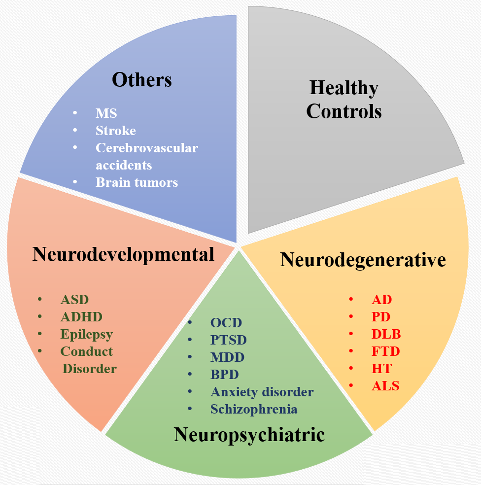
Figure 1
Three major categories of brain disorders (A) Neurodevelopmental; (B) Neuropsychiatric (C) Neurodegenerative, and (D) Others, as well as few examples from each. Abbreviations are: autism spectrum disorder (ASD); attention deficit hyperactivity disorder (ADHD), major depressive disorder (MDD), bipolar disorder (BPD), post traumatic stress disorder, (PTSD), obsessive compulsive disorder (OCD); alzheimer’s disease (AD); parkinson’s disease (PD); dementia with lewy body disease (DLB); fronto temporal disorder (FTD); huntington disease (HT), amyotrophic lateral sclerosis (ALS) and, multiple sclerosis (MS). There are more brain disorders which are not listed above.
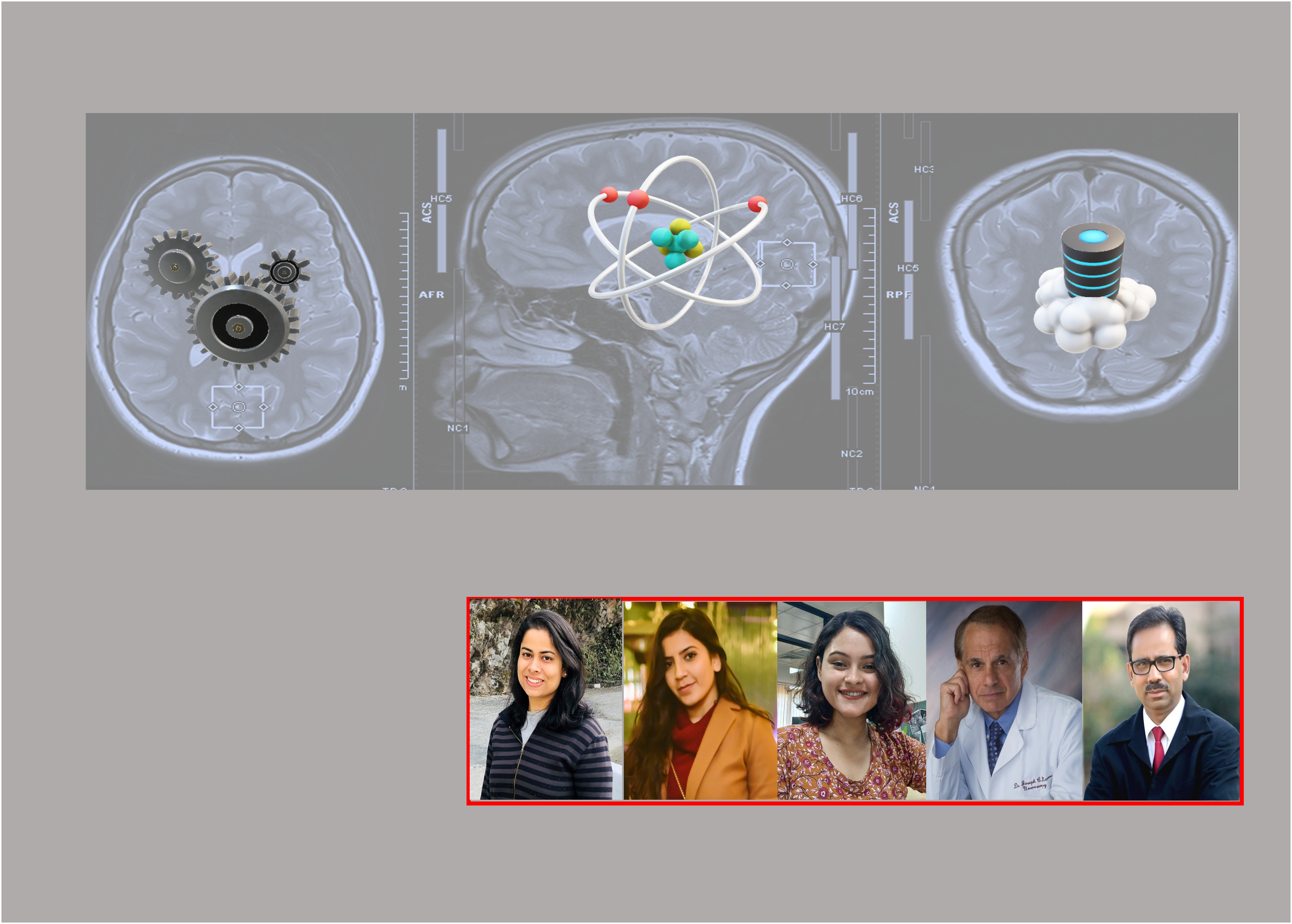
The National Brain Research Center (NBRC) and University of Pittsburgh Medical School (UPMC).
NBRC is the premium research institute in India dedicated solely to studying the brain in health and diseased conditions and is located at the outskirts of the millennium city of Gurugram in the National Capital Region, India. The Neuroimaging and Neurospectroscopy Laboratory (NINS Lab), under the neuroimaging division of NBRC is directed by Prof. Pravat K. Mandal, has its specific purposes to identify early detection biomarkers of Alzheimer's and Parkinson’s disease, development of Indian brain template BRAHMA, and development of various pipelines being used for image analysis and the enrichment of a novel database “SWADESH” platform. The Department of Neurosurgery at the University of Pittsburgh is a world class center for innovative neurosurgery, patient care, and cutting-edge research. Prof. Joseph Maroon, our research collaborative partner is the Heindl Scholar in Neuroscience and the Vice Chairman of neurosurgery, UPMC .
Distinct Features from Various Imaging Modalities
Powerful imaging modalities like magnetic resonance imaging (MRI) provides a three-dimensional structure of the brain and other features (e.g., texture, shape, contrast. etc.). MR Spectroscopy (MRS) provides quantitation of neurochemicals as well as different antioxidants and receptors, noninvasively. The outcome of MRS technique is the determination of brain antioxidant levels, Glutathione (GSH), Gamma-aminobutyric acid (GABA), glutamate and glutamine, neuronal marker, NAA, ATP, PCr, pH, etc. Diffusion tensor imaging (DTI) provides the orientation of directional diffusion of water movement due to tissue microstructural change and provides fractional anisotropy (FA) changes. Quantitatvie susceptibility mapping (QSM) is an advanced MRI technique to evaluate the deposition of heavy metals (e.g., iron, calcification, etc.) arising from dysfunctional molecular processes. Functional MRI (fMRI) is used to measure the changes in blood oxygen-level dependent (BOLD) activities, which is an indirect measurement of deoxyhaemoglobin level modulation due to task-induced neural brain activity and it helps to establish functional connectivity (FC) between various anatomical regions. Single-photon emission computed tomography (SPECT) aids in studying the blood flow to tissues and organs, through the delivery of an intravenous gamma-emitting radioisotope injected into the bloodstream. Quantitative electroencephalography (QEEG) is an imaging modality that assist to detect electrical activity of the brain by measuring the shift in various characteristic signals (α, β, etc.) with the help of sophisticated analysis tools.
A working scheme for database generation involving data analytics.
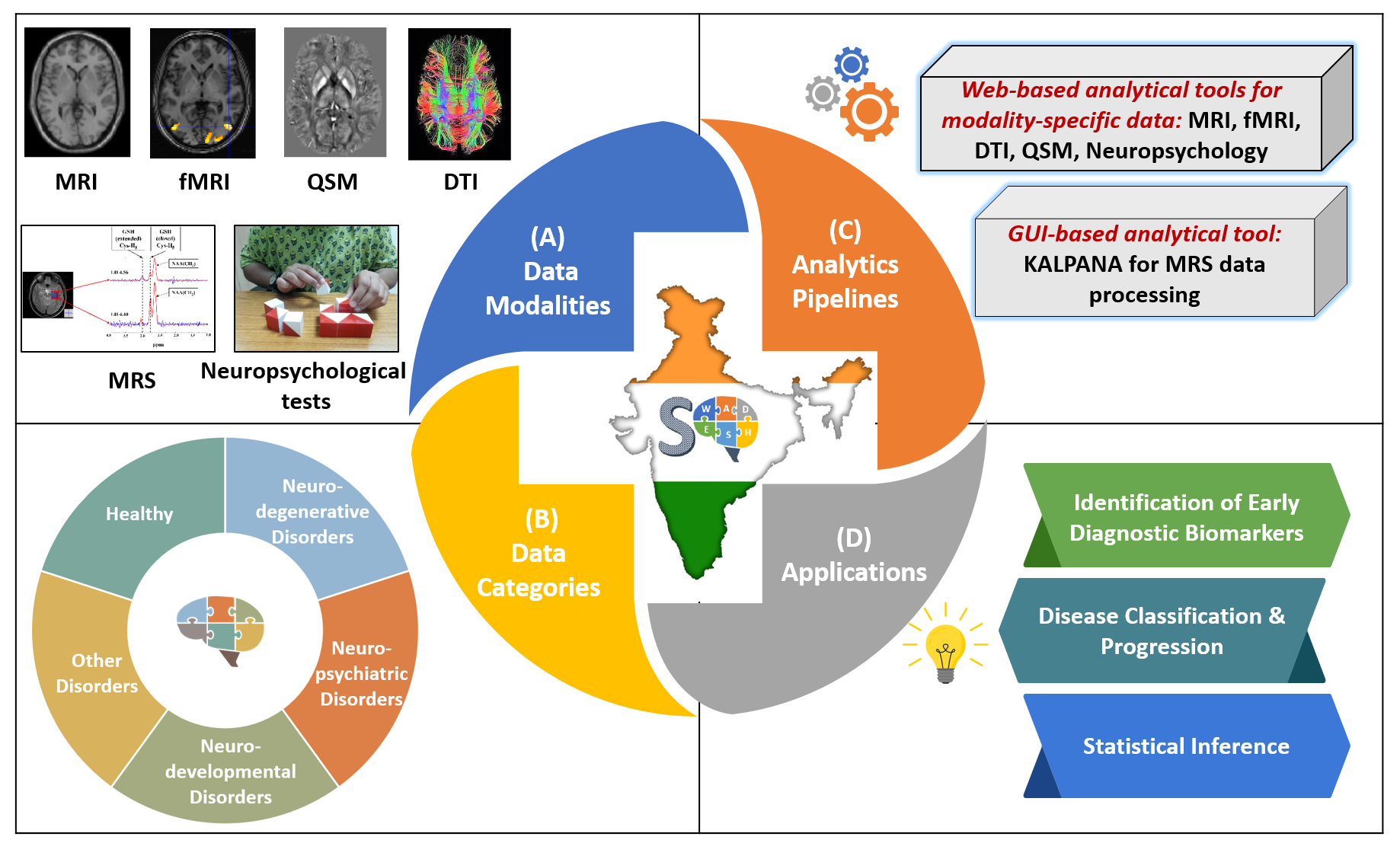
Figure 2
Overview of the SWADESH platform. (a) Data modalities: MRI, MRS, fMRI, DTI, QSM, and neuropsychological data; (b) Data Categories: neurodegenerative, neuropsychiatric, neurodevelopmental, other disorders, and healthy subjects, (c) Analytics pipelines: Web-based analytics pipelines for MRI, fMRI, DTI, QSM and neuropsychological data and KALPANA package for MRS processing. (d) Applications in terms of scope of framework.
Future goal
Even though there are large scale working groups like ADNI for Alzheimer’s disease research, and ENIGMA, an international consortium involving researchers and institutions from each continent for data sharing is urgently required. Neurochemical as well as multi-disease state data and AI powered analysis will enhance identification of the causal process of these disorders. It is our sincere hope that in the coming days, our in-house developed and enriched SWADESH platform will play decisive role for the development of targeted therapeutics.
Acknowledgement:
We (Yashika Arora, Avantika Samkaria, Rimil Guha Roy, Joseph Maroon & Pravat K Mandal) dedicate this blog to the healthy control participants, patients who participated in various clinical studies in last 15 years at NINS lab, NBRC and their family members for cooperation.
Please sign in or register for FREE
If you are a registered user on Research Communities by Springer Nature, please sign in