Painting the human body with light and sound
Published in Healthcare & Nursing and Bioengineering & Biotechnology
When we first began discussing this project several years ago, our goal was both simple and ambitious: could we find a practical way to combine the strengths of ultrasound and optical imaging to visualize the human body more completely, without adding complexity to the clinical workflow?
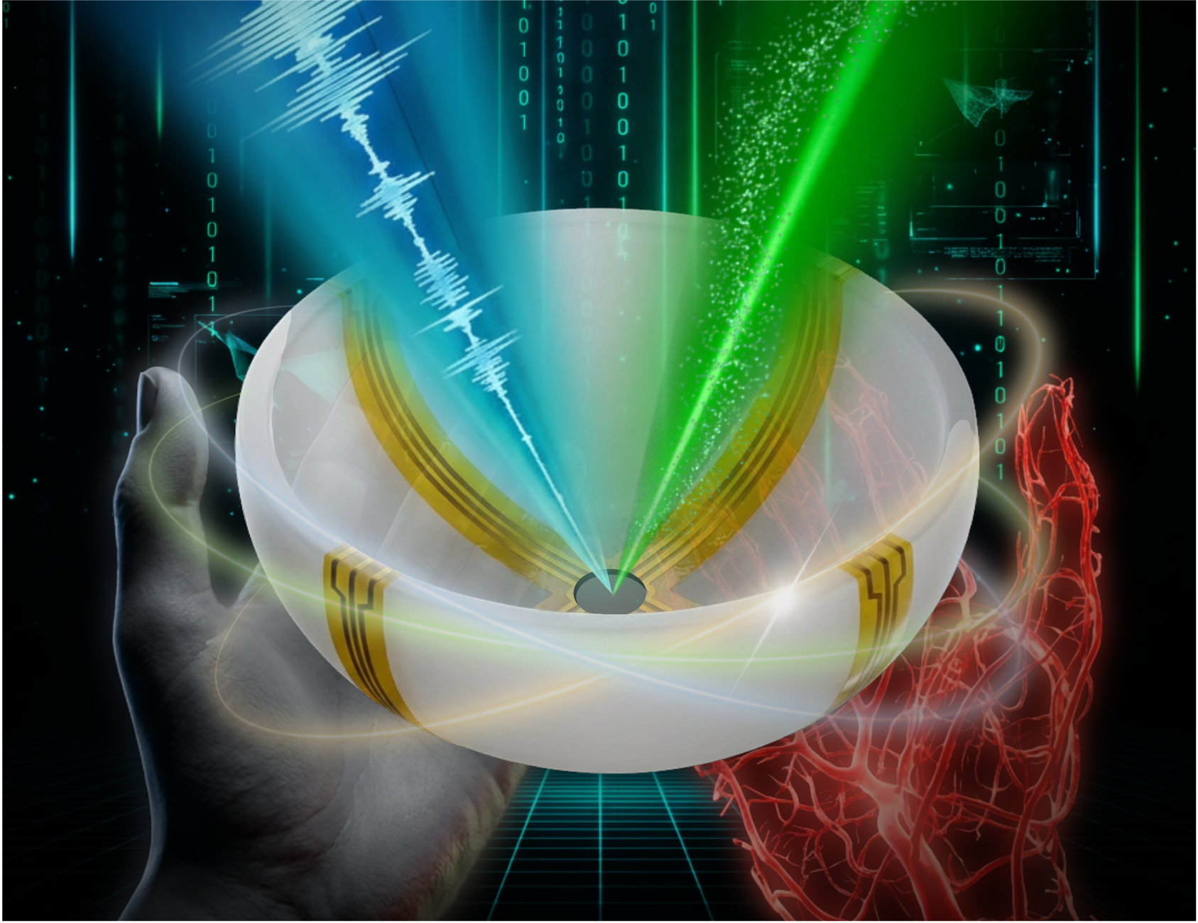
This question eventually led to our recent paper in Nature Biomedical Engineering, “Rotational ultrasound and photoacoustic tomography of the human body.” In this work, we introduce a hybrid imaging platform—RUS-PAT—that integrates rotational ultrasound tomography (RUST) and photoacoustic tomography (PAT) to enable quasi-simultaneous three-dimensional imaging of tissue structure and vasculature in humans.
Why combine ultrasound and photoacoustics?
Medical imaging remains a field of trade-offs. Ultrasound is widely used, affordable, and safe, but its contrast is primarily structural. Optical methods, on the other hand, provide rich sensitivity to blood and molecular absorbers, yet suffer from limited penetration and strong scattering in tissue.
Photoacoustic tomography offers an elegant bridge between these worlds: optical absorption generates ultrasound waves that can be detected deep inside tissue. However, most photoacoustic systems lack quantitative structural context, while conventional ultrasound tomography alone cannot reveal large–field-of-view vascular or functional information.
We wondered whether it was possible to integrate both modalities into a single rotating system that could acquire complementary datasets in one scan, without sacrificing speed or field of view.
Building a practical hybrid system
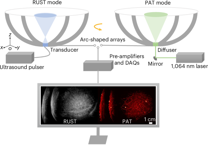
The solution we developed uses a rotating arc-shaped ultrasound transducer array combined with synchronized pulsed laser excitation and a single source ultrasound transducer. During successive rotational scans, the system first collects ultrasound tomography data and then photoacoustic signals, allowing us to reconstruct volumetric images of tissue morphology and vasculature in the same coordinate frame.
With this approach, RUS-PAT achieves a field of view of approximately 10 cm, sub-millimetre isotropic resolution, and scan times of about ten seconds. We demonstrated three-dimensional dual-contrast imaging across multiple anatomical regions, including the head (brain), breast, hand, and foot, without ionizing radiation or exogenous contrast agents.
A recent Caltech news feature described this concept as “bringing optical color to ultrasound,” which nicely captures the motivation behind the work. By adding optical absorption contrast to conventional ultrasound tomography, the system reveals vascular networks and functional information that would otherwise remain invisible.
Toward clinical translation
From the beginning, we emphasized simplicity and clinical relevance. Rather than building a highly specialized laboratory instrument, we designed RUS-PAT as a modular platform that can interface with existing photoacoustic or ultrasound hardware. Our validation experiments included both tissue phantoms and in vivo human imaging, demonstrating stable performance across a range of anatomies.
We believe this hybrid approach may complement existing imaging workflows in applications such as vascular assessment, breast imaging, musculoskeletal evaluation, and potentially transcranial studies.
Hybrid imaging with light and sound continues to evolve, and many exciting directions remain. Future developments may include extending the platform to dual-modality human brain imaging and exploring applications in breast cancer diagnosis and monitoring.
This project was the result of close collaboration between engineers, physicists, and clinicians at Caltech and USC. I am deeply grateful to all collaborators and mentors who contributed their ideas, expertise, and patience throughout this journey.
We hope that RUS-PAT will inspire further efforts to develop practical multimodal imaging systems—and help bring richer, more informative imaging tools into everyday clinical practice.
Cite this article
Zhang, Y., Na, S., Russin, J.J. et al. Rotational ultrasound and photoacoustic tomography of the human body. Nat. Biomed. Eng (2026). https://doi.org/10.1038/s41551-025-01603-5
Follow the Topic
-
Nature Biomedical Engineering
This journal aspires to become the most prominent publishing venue in biomedical engineering by bringing together the most important advances in the discipline, enhancing their visibility, and providing overviews of the state of the art in each field.

Related Collections
With Collections, you can get published faster and increase your visibility.
Implantable wireless communication technologies
Publishing Model: Hybrid
Deadline: Nov 28, 2026
Medical Ultrasound: Emerging Techniques and Applications
Publishing Model: Hybrid
Deadline: Jan 29, 2027

Please sign in or register for FREE
If you are a registered user on Research Communities by Springer Nature, please sign in