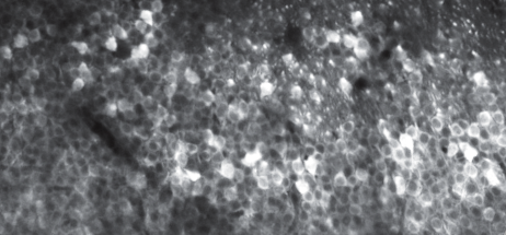




Calcium imaging is an amazing method that allows us to look deep into the brain of a living animal and observe its activity at the level of single cells. Seeing individual cells firing in a living animal is an experience that I find still fascinating, even after >10 years in neuroscience.
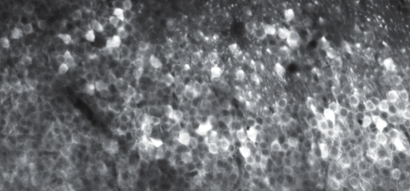
However, the beauty of the videos hides some problems with calcium imaging data. When you look closely, it becomes apparent that the calcium imaging signals are not so easy to interpret and how they relate to neuronal activity, that is, action potentials or "spikes".
Over the past two decades, many studies have claimed that they could use calcium imaging to resolve individual spikes, which would be an important achievement. This is an important point: if calcium signals could reliably report individual spikes, they would provide a calibrated and directly meaningful way to read neural activity at scale. But if not, then the recorded signals would be ill-defined "activity patterns" that are difficult to connect to the fundamental unit of information processing in the brain, the spike. However, after careful inspection, these claims were refuted or found to apply to only a subset of cells or only very favorable and unrealistic conditions.
That's why I was highly skeptical when in November 2021 a new study appeared as a preprint (later published in Nature). The preprint claimed to have found a calcium indicator that reliably reports individual spikes. As an expert in this matter, I carefully read the paper and remained skeptical.
My goal was not to confirm the claim, but to disprove it. I set out to show that for this new indicator - named GCaMP8 - detection of single spikes was not reliably possible. First, I downloaded the large dataset with simultaneous calcium imaging and electrophysiological recordings, which was generously shared by the scientist who had acquired it, Márton Rózsa.
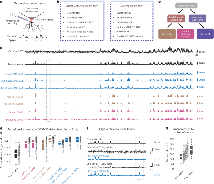
I worked on this project over the years, carefully inspecting the raw data, but also performing many complicated analyses, while testing the indicator also in the lab for ongoing experiments. Over time, the originally small project idea became larger and expanded. I performed systematic analyses using different algorithms for "spike inference" (to analyze whether a spike was present or not), using both algorithms that I wrote myself and algorithms from others. And I supervised a Master's student (Xusheng "Felix" Fang) on this topic, and his results made a relatively small but interesting contribution to the overall analyses.
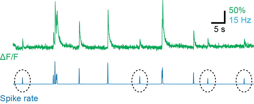
In the end, we found something I did not expect: two variants of this calcium indicator (GCaMP8m and GCaMP8s) were indeed capable of reliably detecting single spikes! This was opposite to my expectations - but good news for the scientific community!
In addition, we made another interesting observation: the ability to detect single spikes was not primarily the consequence of one indicator being brighter than others, or showing a higher dynamic range, which would both enable better detection. Instead, it was a higher "linearity". What does this mean? Nonlinear behavior of a calcium indicator in this case means that the indicator responds very weakly for a single spike but then very strongly for a burst of 2 or 3 spikes.
A nonlinear calcium indicator behaves like a contrast enhancer, which we use in imaging processing software to make our pictures look nicer: we make the transfer curve nonlinear (sigmoidal), which increases the contrast but also loses some of the finer details.

In our work, we showed that this nonlinear behavior was exactly how earlier calcium indicators worked; the newer calcium indicators (GCaMP8s and GCaMP8m) did not exhibit this nonlinear behavior for individual spikes, enabling us to detect single spikes (which are "the finer details") with calcium imaging.
Of course, this work is not the final word about calcium imaging and how to detect action potentials with it. There is still room for algorithms to improve on our methods for "spike inference", and for indicators that are more linear across a larger range of activity. In addition, these findings need to be validated across cell types and brain regions.
Overall, calcium imaging lets us watch the brain in action, but only if we understand what we are seeing. Our work brings us a step closer to reading those signals correctly. And this is important, because, as we write in our own paper, "the practical value of new tools depends not only on their quality but also on rigorous validation and clear guidance for use."

I am Peter Rupprecht, a neuroscientist and group leader based in Switzerland. Please find my CV here.
Since March 2023, I’m leading a junior research group in the lab of Fritjof Helmchen at the Brain Research Institute in Zürich/Switzerland. We use and develop two-photon microscopy for calcium imaging in hippocampus from both neurons and astrocytes, large-scale imaging of cleared brain tissue, closed-loop behavioral paradigms, and single-cell perturbations in vivo. Key components of our work are careful experimental design as well as in-depth data analysis rather than large-scale experiments or screens, with the goal to understand individual cells and how they integrate signals from local and brain-wide circuits.
Follow the Topic
-
Nature Methods
This journal is a forum for the publication of novel methods and significant improvements to tried-and-tested basic research techniques in the life sciences.

Related Collections
With Collections, you can get published faster and increase your visibility.
Methods development in Cryo-ET and in situ structural determination
Publishing Model: Hybrid
Deadline: Oct 30, 2026

