Shaping of crystals at the nanoscale by anisotropic capillary waves
Published in Electrical & Electronic Engineering

Great as the differences between underwater corals and nanoparticle superlattices, they are both the crystalline assemblies from nanosized building blocks, and they share the same importance of crystal shapes on their properties—whether the color and colony of life residing upon them or the wavelength and modes of light-matter interactions. However, despite the rapid progress in nanotechnology over the last few decades, understandings remain limited on how the functional nanosized building blocks assemble into highly ordered structures against stochastic Brownian motions in a liquid medium. Filling this gap requires high spatiotemporal resolution imaging of dynamics in a liquid environment, which has been challenging. In this work, the emergent technique of liquid-phase transmission electron microscopy (TEM) was used to overcome this challenge and capture the emergence of a faceted crystal shape from individual nanosized building blocks. Anisotropic capillary waves were imaged directly at nanometer and sub-second resolution to back out the facet-defined surface energy of the crystal.

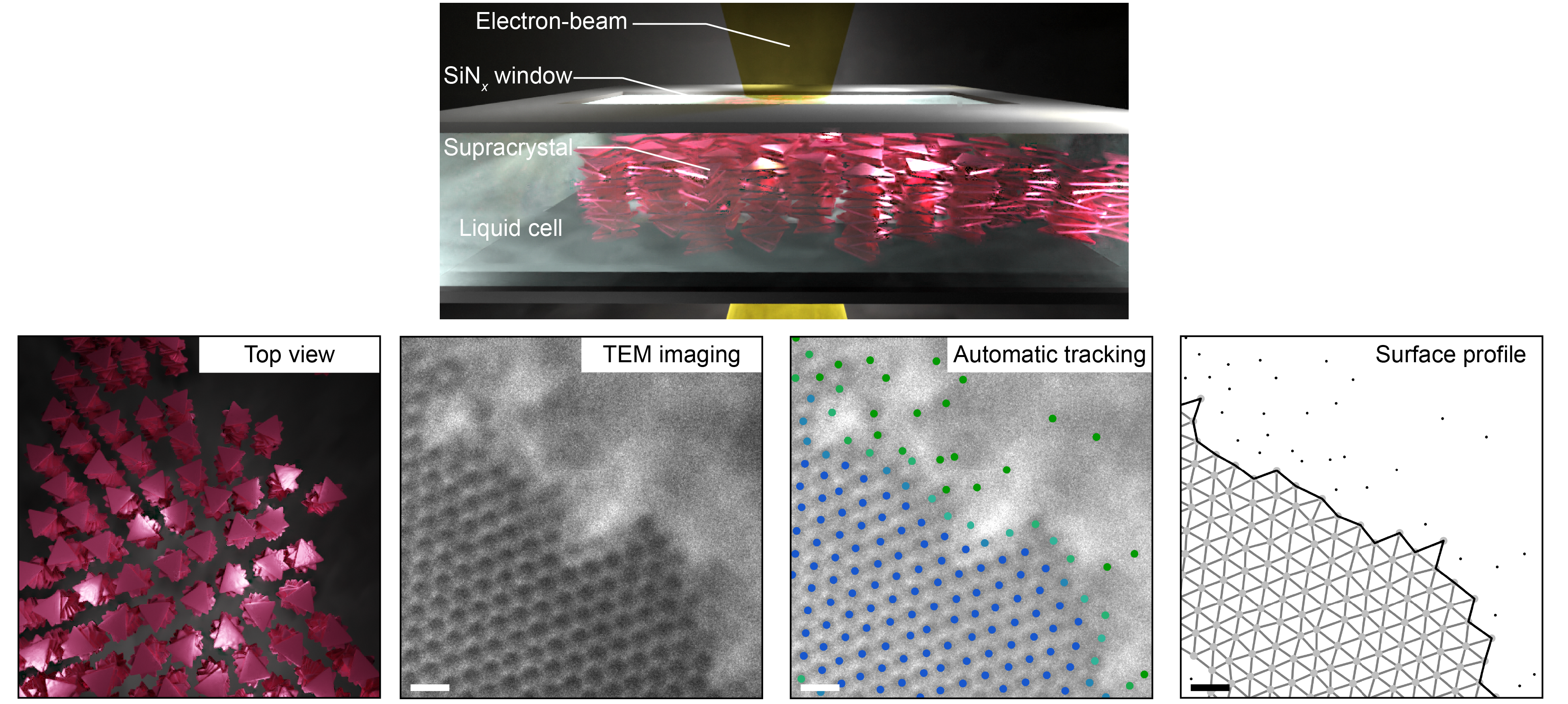
Figure 1. A supracrystal–suspension interface visualized in real-time by liquid-phase transmission electron microscopy (TEM). (Top) Schematic showing the configuration of liquid-phase TEM. (Bottom) Illustration of how liquid-phase TEM is combined with automatic tracking and structural characterization to extract the surface profile of a supracrystal. Scale bars: 200 nm.
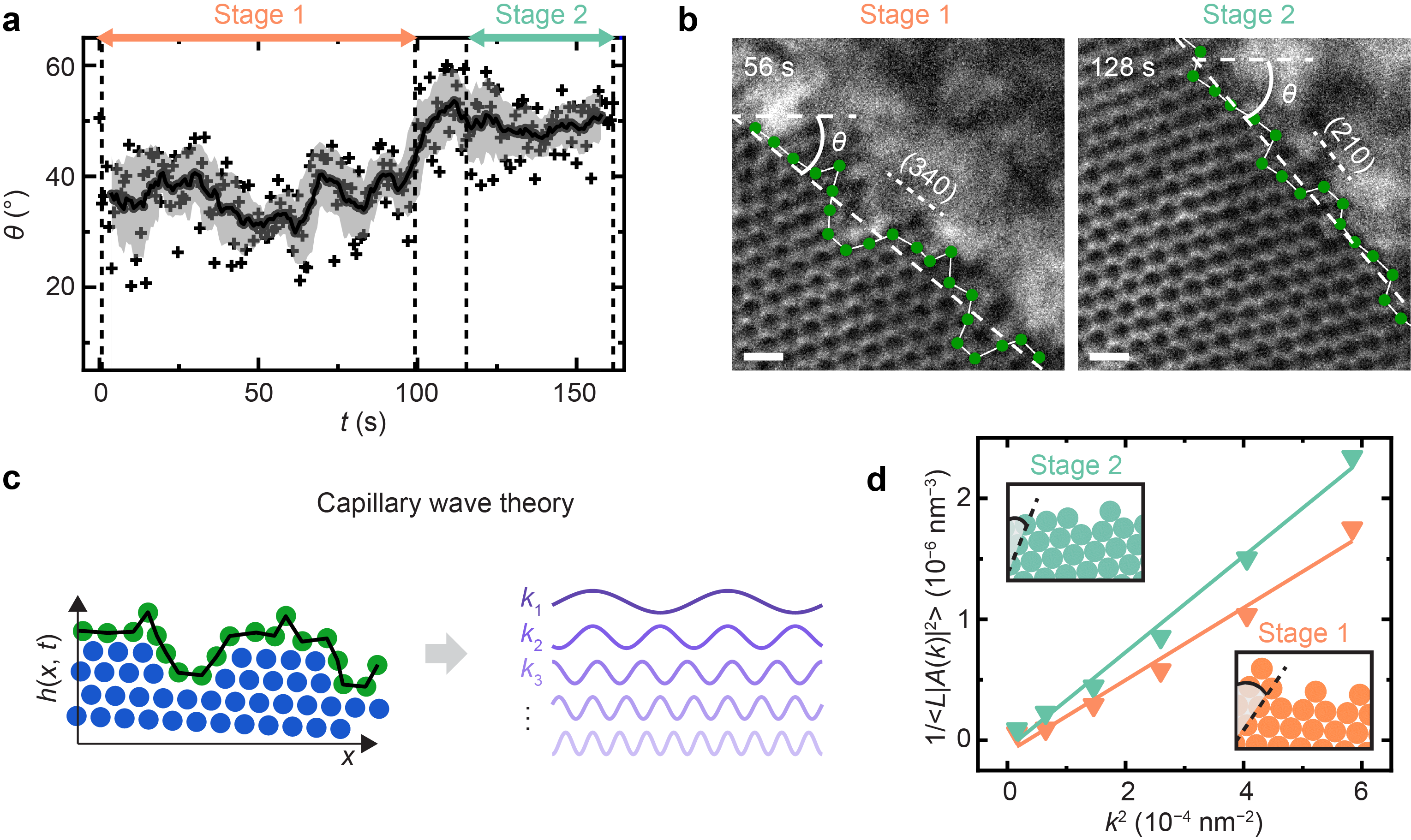
We integrate low-dose liquid-phase TEM imaging, single nanoparticle tracking, and quantitative structural analysis to identify and monitor real-space surface profiles of a growing nanoparticle supracrystal (Fig. 1). Interestingly, the supracrystal shifts its surface orientation within our observation time window as it grows from continuous attachments of nanoparticles, presenting two temporal stages (Fig. 2a). By closely looking into the surface profiles, we were reminded of the long-standing capillary wave theory by their dynamic fluctuations. Capillary wave theory treats surfaces or interfaces as superimposed waves and the equilibrium profile is a tradeoff between the surface energy and thermal fluctuation. We first demonstrate the theory’s applicability to the nanoscale and to the surface of a crystal (instead of the more disordered liquid phase), suggesting the randomness of Brownian motions still plays a major role in defining the surface. We are then able to measure a series of otherwise inaccessible physical parameters, including surface roughness, correlation length, interfacial stiffness and mobility, for different supracrystal facets. Interestingly, the interfacial properties we quantify in the experiments are anisotropic and the supracrystal grows towards exhibiting flatter surfaces with lower surface energy, consistent with the Wulff construction rule proposed to explain the equilibrium shape of a crystal (Fig. 2b‒c).

Figure 2: Shaping of crystals at the nanoscale by anisotropic capillary waves. (a) Temporal evolution of the crystal surface orientation shows two stages. (b) Typical liquid-phase TEM images for stage 1 and stage 2 highlighting the surface orientation shift. Scale bars: 200 nm. (c) Schematic illustrating the decomposition of the surface profile into superimposed waves with different wave vectors. (d) Anisotropic interfacial stiffness measured for different facets based on capillary wave theory.
Our capability to measure facet-dependent interfacial energies and our demonstration on their role in shaping the supracrystals can advance crystal design at the nanoscale. For example, one can control the interfacial stiffness and crystal habit by utilizing the toolkits of both intrinsic parameters of nanoparticle shape and surface chemistry as well as extrinsic parameters such as temperature, pH and ionic strength. Our approach can elucidate the fundamental role of these parameters in guiding crystallization. Broadly, our method can be applied to study other nanoscale fluctuations as liquid-phase TEM becomes more compatible with biological samples (e.g., fusion of lipid vehicles) and with applying external field (e.g., dendrite formation at the electrolyte–solid interface).
This work was recently published in Nature Communications:
Imaging How Thermal Capillary Waves and Anisotropic Interfacial Stiffness Shape Nanoparticle Supracrystals
Zihao Ou, Lehan Yao, Hyosung An, Bonan Shen and Qian Chen
Nature Communications, 11, 4555 (2020): https://www.nature.com/articles/s41467-020-18363-2
Follow the Topic
-
Nature Communications

An open access, multidisciplinary journal dedicated to publishing high-quality research in all areas of the biological, health, physical, chemical and Earth sciences.
Related Collections
With Collections, you can get published faster and increase your visibility.
Women's Health
Publishing Model: Hybrid
Deadline: Ongoing
Advances in neurodegenerative diseases
Publishing Model: Hybrid
Deadline: Mar 24, 2026


Please sign in or register for FREE
If you are a registered user on Research Communities by Springer Nature, please sign in