The Alpha Rhythm: From the Origins of EEG to Brain Aging and Health Equity
Published in Neuroscience



Just over a century ago, Hans Berger published the first study of the human resting-state electroencephalogram (rsEEG), opening an unprecedented path for the study of brain dynamics. Despite major advances in neuroimaging since those early observations, the alpha rhythm — 8-12 Hz oscillations that constitute the hallmark signature of rsEEG — has remained a powerful marker of brain function. As people age, the alpha rhythm shows significant slowing and reduced power. In Alzheimer’s disease, the most prevalent neurodegenerative disease in older adults, these alterations become pronounced and are closely associated with cognitive decline.
Aging, however, is not only the passage of time. It involves the gradual decline of physiological functions that support survival, adaptation, and reproduction. Because this decline varies across organs and organ systems, chronological age offers a picture of health that is complemented by biological age, which informs how old a body system appears in anatomical or functional terms. Brain clocks bring this idea into neuroscience. Using machine-learning models, they estimate a person’s brain age from brain data and compare it with their chronological age. This difference, known as the brain age gap (BAG), can indicate whether the brain appears older or younger than expected. A positive BAG has been linked to accelerated brain aging and neurodegenerative conditions. Consequently, the BAG has emerged as a promising indicator of brain health.
Brain health reflects the interplay between biology and environment. Therefore, a major challenge in this area is developing global models that capture shared mechanisms across populations while remaining sensitive to geographic, socioeconomic, and cultural contexts. Building such models requires brain measures that are both biologically meaningful and accessible across different research and healthcare settings. EEG is especially well-suited to this purpose because it directly captures ongoing brain electrical activity with high temporal resolution and is portable, relatively low-cost, and easy to deploy.
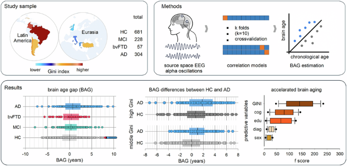
To formulate our research question, we intentionally returned to the foundations of EEG: alpha activity. We asked whether this EEG rhythm could serve as a reliable measure of brain aging that is meaningful in clinical settings and sensitive to population diversity and social disparities. Our work was conducted within the EuroLAD-EEG Consortium, a collaborative platform for dementia research that integrates rsEEG, clinical, and sociodemographic data from participants across countries in the Global South and the Global North. We analyzed data from 1,228 participants across 10 countries with varying levels of structural inequality. The sample included healthy participants, those with mild cognitive impairment, and patients with Alzheimer’s disease or behavioral variant frontotemporal dementia. We trained machine-learning models to predict brain age from EEG source-space alpha activity and calculated BAG for each participant. To build these models, we used both canonical alpha bands and individualized alpha features.
The results showed that alpha-based BAG increased along the neurodegenerative continuum. Healthy participants had the lowest BAG, whereas individuals with mild cognitive impairment and dementia showed gradually higher values. In other words, the alpha activity-based brain clock captured deviations from the expected aging trajectories. The brain regions most relevant to the model were also biologically meaningful. Occipital, parietal, cingulate, and hippocampal regions contributed substantially to brain age prediction. These areas are closely linked to posterior alpha rhythms, attention, memory networks, and vulnerability to neurodegeneration. The most striking finding was that structural inequality mattered. Among healthy individuals, BAG increased with country-level income inequality. When we examined factors distinguishing participants with higher versus lower BAG, structural inequality emerged as the strongest predictor, surpassing cognition, education, sex, and even clinical diagnosis in some analyses. This indicates that brain aging is a physiological process shaped by the broader social environments in which individuals live, and that macrosocial conditions leave identifiable traces in brain dynamics, reflected in an easy-to-record, well-understood, and clinically familiar EEG descriptor: alpha waves.
Alpha-based EEG brain clocks and, more generally, EEG-based brain-age models can enrich the toolkit for assessing brain health. Alongside MRI, molecular biomarkers, and comprehensive clinical evaluations, they offer a functional, interpretable, and scalable measure of brain aging and neurodegeneration. This is important for advancing health equity and universal health coverage, as many communities still have limited access to advanced neuroimaging and biomarker testing. In this context, EEG-based tools could improve access to brain health assessment, support large-scale screening and ongoing monitoring, and foster more inclusive research participation in diverse and underserved populations.
Therefore, behind this paper is the conviction that a global approach to brain health, aimed at serving diverse populations worldwide, must rely on accurate, interpretable tools that can be implemented in underserved settings. Alpha activity embodies this possibility, connecting EEG’s historical foundations with current challenges in aging, dementia, and health equity. We hope that collaborative, transdisciplinary efforts, such as those that made this study possible, will continue to deepen our understanding of brain health and, in doing so, help improve the lives of underserved populations around the world.
Follow the Topic
-
Communications Biology
An open access journal from Nature Portfolio publishing high-quality research, reviews and commentary in all areas of the biological sciences, representing significant advances and bringing new biological insight to a specialized area of research.

Related Collections
With Collections, you can get published faster and increase your visibility.
Healthy Aging
Publishing Model: Open Access
Deadline: Dec 31, 2026
DNA repair and human disease
Publishing Model: Hybrid
Deadline: Oct 31, 2026